Predictors of Therapy Response in Chronic Aphasia: Building a Foundation for Personalized Aphasia Therapy

Article information

J Stroke. 2022;24(2):189-206
Publication date (electronic) : 2022 May 31
doi : https://doi.org/10.5853/jos.2022.01102
aCenter for the Study of Aphasia Recovery, University of South Carolina, Columbia, SC, USA
bDepartment of Communication Sciences and Disorders, University of South Carolina, Columbia, SC, USA
cDepartment of Psychology, University of South Carolina, Columbia, SC, USA
Correspondence: Sigfus Kristinsson Department of Communication Sciences and Disorders, University of South Carolina, 915 Greene Street, Columbia, SC 29209, USA Tel: +1-803-553-4689 Fax: +1-803-777-9547 E-mail: sigfus@email.sc.edu
Received 2022 May 31; Revised 2022 April 20; Accepted 2022 April 21.

Abstract

Chronic aphasia, a devastating impairment of language, affects up to a third of stroke survivors. Speech and language therapy has consistently been shown to improve language function in prior clinical trials, but few clinicially applicable predictors of individual therapy response have been identified to date. Consequently, clinicians struggle substantially with prognostication in the clinical management of aphasia. A rising prevalence of aphasia, in particular in younger populations, has emphasized the increasing demand for a personalized approach to aphasia therapy, that is, therapy aimed at maximizing language recovery of each individual with reference to evidence-based clinical recommendations. In this narrative review, we discuss the current state of the literature with respect to commonly studied predictors of therapy response in aphasia. In particular, we focus our discussion on biographical, neuropsychological, and neurobiological predictors, and emphasize limitations of the literature, summarize consistent findings, and consider how the research field can better support the development of personalized aphasia therapy. In conclusion, a review of the literature indicates that future research efforts should aim to recruit larger samples of people with aphasia, including by establishing multisite aphasia research centers.

Introduction

Aphasia is a devastating language disorder most commonly resulting from a cortical lesion to the perisylvian region of the language-dominant hemisphere [1,2]. Despite a general lack of public knowledge [3], chronic aphasia (≥6 months post-onset) is not an uncommon disorder, affecting up to a third of stroke survivors [4]. Behavioral speech and language therapy (SLT) has been shown to be an efficacious approach to improve language function in persons with aphasia (PWA) as a group [5,6], and remains a mainstay in the clinical management of aphasia [7]. Nonetheless, there is notorious and unexplained heterogeneity in therapeutic effects at the individual level [8-10]. Critically, prior efforts to identify individual predictors of therapy response [11-13] have not yet managed to reliably answer two fundamental questions: (1) who responds to SLT and (2) to what degree? Meanwhile, clinicians who routinely administer SLT struggle with personalized therapy planning and prognostication [14].

More specifically, clinicians are presently unable to determine with a consistent degree of confidence whether and to what degree any given PWA will respond to therapy based on pre-therapy individual characteristics. To this end, addressing questions (1) and (2) above is a crucial prerequisite to facilitate the development of personalized aphasia therapy (PAT), i.e., therapy tailored to maximize each individual’s potential language recovery with reference to evidence-based clinical guidelines.

Innovative technological developments and advances in therapeutic approaches in the literature have increasingly emphasized the pressing demand for personalized solutions in aphasia therapy [15,16]. However, few generalizable and clinically applicable predictors of therapy response have been identified to date. The primary cause likely stems from the fact that the relevant literature is predominated with single-subject and small group studies [13,17]. While single-subject and small group study designs are well-suited to acquire detailed accounts of the experiences of individuals with specific characteristics, they are, by definition, not intended to reflect population parameters. From a statistical perspective, the issue of individual variability in therapy response is substantially exacerbated due to small sample sizes, which leaves most prior studies with severely limited statistical power to detect causal relationships that can be leveraged to guide PAT at the population level [18,19]. Epidemiological trends indicating a rising prevalence of aphasia [20] and, as a result, a growing societal burden of aphasia [21], have rendered research aiming to improve clinical management of aphasia an immediate public health priority.

In the interest of facilitating the development of PAT, this narrative review offers a comprehensive overview of the current state of the literature on personalized predictors of therapy response in chronic aphasia. We restrict our focus to chronic aphasia as recent large-scale randomized controlled trials have failed to demonstrate robust therapeutic effects beyond spontaneous recovery when therapy was commenced in the acute (≤2 weeks of stroke onset) phase of recovery [22-24]. Specifically, we emphasize recent studies that have explicitly examined the association between common biographical, neuropsychological, or neurobiological variables, and response to impairment-based SLT. Given the limitations of the literature, indirect evidence, such as cross-sectional studies, studies of spontaneous aphasia recovery, and other derived sources are similarly considered as applicable. We conclude by discussing how clinical prognostication can be improved through future research endeavors.

Predictors of treated recovery in aphasia

There is an unequivocal and obvious benefit of identifying robust predictors of therapy response in aphasia. Nevertheless, few comprehensive studies have been carried out in pursuit of this goal. One potential reason is the cost of conducting large-scale therapy studies in aphasia. The cost associated with participant recruitment, administration of therapy, collection of an extensive dataset, and multiple magnetic resonance imaging (MRI) scanning sessions per participant is high, and accomplishing studies of this scale requires both an interdisciplinary team of professionals and tangible resources. Notwithstanding, a few studies have directly examined predictors of therapeutic effects in samples of varying sizes [25]. The evidence amassed through these studies is discussed in what follows.

Biographical predictors

Age

The brain’s capacity for cognitive processing decreases gradually in normal aging [26]. Granted that language restoration relies on functional and/or structural plasticity and numerous findings showing a steady decrease in brain plasticity with age [27,28], intuition might suggest that older individuals are less likely to show favorable language recovery. In line with this view, several studies have suggested that younger age might mediate positive outcomes in the acute recovery phase [29-35]. For instance, Ali et al. [29] (2021) found younger age (<55 years) to be the strongest predictor of recovery in a large sample of PWA. However, this finding has not been consistently replicated in similar studies [36-40]. One potential reason for this discrepancy is that the relationship between age and recovery may be confounded by stroke age; aphasia is more likely to emerge as a consequence of stroke in older individuals [39-42] and older individuals generally present with more severe aphasia (i.e., fluent aphasia as opposed to nonfluent) [42-45].

Several aphasia therapy studies have observed greater therapy-induced improvements in younger compared to older individuals [46-50]. Recent research in our lab supports these findings. A retrospective study examining the effects of age on outcomes in several prior therapy studies conducted in our lab found a complex relationship between age and outcomes [51]. Johnson et al. [51] reported multicollinearity between age and variables such as sex and education, whereas the independent effects of age varied across studies. By contrast, a number of other aphasia therapy studies have failed to find a consistent relationship between age and therapy success [52-55]. Correspondingly, recent qualitative literature reviews have consistently described the relationship between age and language function in aphasia as equivocal [2,37,56]. Thus, while further research is warranted, any given study aiming to predict therapy response in aphasia should examine the effects of age on the known effect of age on brain repair mechanisms.

Sex

A long-held view that postulates sex differences in language processing has motivated research on the effects of sex on aphasia recovery [57,58]. In their seminal study, Shaywitz et al. [58] noted strongly left-lateralized frontal activation in males, whereas more diffuse neural systems were engaged in females bilaterally in response to a language task. Consistent with the notion that females have more flexibility to recover from left hemisphere (LH) stroke due to bilateral engagement, some studies have noted better recovery of language function in females [29,59,60]. However, most studies have not found differences in language recovery depending on sex [36-39,55,61]. Relatively better recovery in males has even been reported [32]. Therefore, at present, there is no conclusive evidence to suggest that sex is a determining factor of therapy-induced language recovery.

Handedness

Differences in language recovery based on handedness are grounded in theories of language lateralization, similar to theorized differences based on sex. Children born left-handed have been shown to exhibit more bilateral cortical representation of language compared to right-handed children (15%–33% vs. 7%–9%, respectively) [62], although comparably pronounced differences have not been observed in adults [62-64]. Prior studies have not provided conclusive support for the notion that left-handed individuals have greater capacity for language recovery due to greater bilateral representation of language [2,37,39].

Education

Research examining the relationship between education and language recovery draw from the idea that a greater degree of education may be an indication of relatively preserved cognitive reserve after brain damage. While few studies have directly assessed the predictive value of education for therapy outcomes, conflicting results have been reported regarding the effects of education on language recovery in general [31,65-68]. Connor et al. [69] and Hillis and Tippett [65] both reported an association between education and aphasia severity in the chronic phase of recovery, but not with extent of language recovery. The authors noted that education might serve as a proxy variable for a plethora of other factors such as discipline or determination, cognitive reserve, economic resources, healthy lifestyle, literacy level, socio-economic status, occupation, and access to healthcare [70]. Thus, it remains to be determined whether education independently influences response to therapy.

Time post-stroke

Time post-stroke (TPS) is an obvious and crucial determinant of early spontaneous recovery of language function, as the recovery trajectory is steepest in the 1st weeks through 6 to 12 months following stroke onset [39,59,71]. In the chronic phase, TPS does not seem to be related to response to therapy in PWA [53,54,72,73]. Moss and Nicholas [73] offered a comprehensive review of the effects of TPS on therapy outcomes in individuals beyond 1-year post-onset. In short, the authors found no correlation between TPS at which therapy was initiated with response to therapy [73].

Psychosocial factors

The literature on the effects of psychosocial factors such as social support and mood on therapy response is scarce, but a handful of studies have found that social support and mood contribute to individuals’ quality of life and sense of autonomy [74-76]. These findings have led some researchers to argue that strong social networks and motivation facilitate progress in therapy [16,77]. Consequently, functional communication and community support therapy approaches generally focus on strengthening social relationships and improving mood with the intention of enhancing individuals’ sense of autonomy. The Life Participation Approach to Aphasia (LPAA) philosophy, which aims to empower PWA to actively participate in their rehabilitation and to engage in daily activities of interest, is particularly relevant in this respect [78]. Group-based therapy approaches likewise aim to combat the social isolation commonly experienced by PWA [79-81]. Furthermore, various language therapy approaches have been found to improve mood in PWA [82-84]. Therefore, psychosocial factors may contribute to the likelihood of positive outcomes following therapy, although further research on the precise psychosocial predictors is necessary.

Aphasia type

Aphasia type is dependent on factors such as lesion size and location, and by extension higly correlated with aphasia severity. Nonetheless, aphasia classification has been investigated in relation to clinical outcomes [8,40,59]. In terms of therapy response, however, the independent predictive value of aphasia type remains unclear. Individuals with Broca’s aphasia may have positive prognosis in the 1st year following stroke [85], whereas in the chronic phase, individuals with fluent aphasia subtypes (e.g., anomic and conduction aphasia) have been shown to respond better to therapy than nonfluent individuals [8,86]. For example, Kristinsson et al. [8] observed a larger overall therapy effect in fluent compared to nonfluent individuals. These findings suggest that aphasia type can inform prognostication in aphasia therapy, although the potential confounding effects of aphasia severity, lesion size, and location need to be taken into consideration.

Summary of biographical predictors

In terms of biographical predictors, limited generalizable knowledge has accumulated in the literature. Prior studies suggest a complex and potentially confounding relationship between age and aphasia type with therapy success. Time since stroke-onset does not seem to negatively affect therapy response in chronic aphasia, and sex and handedness similarly do not seem to affect therapy response. On the other hand, social support and motivation are potentially favorable attributes, but further research where these predictors are better defined is needed. Similarly, the independent predictive value of education remains to be determined. In conclusion, systematic evaluation of biographical predictors of therapy outcomes is needed before this knowledge can be applied in clinical practice.

Neuropsychological predictors

Aphasia severity

The initial severity of language deficits is commonly recognized as the most consistent predictor of aphasia recovery [2,70]. Aphasia severity is strongly associated with spontaneous recovery, with increased rate of complete recovery observed in individuals with milder symptoms [33,34,39,66,67,87]. Correspondingly, aphasia severity is generally considered the single strongest predictor of response to impairment-based therapy [16,17,48,59]. Performance on confrontation naming tasks, as a proxy measure for aphasia severity, has similarly emerged as the strongest predictor of response to anomia therapy [12,55,88,89]. For instance, Seniów et al. [55] found that severity of anomia prior to therapy was highly correlated with all post-therapy language outcomes in a relatively large sample of 78 participants with chronic aphasia.

Despite the broad consensus in the literature, the precise nature of the relationship between aphasia severity and therapy outcome has yet to be fully mapped out. While some researchers have found a relative advantage for individuals with milder aphasia [52,88], others have observed an advantage for individuals with more severe aphasia [90], and some have reported mixed results [54]. Recent work from our lab offers a potential explanation for this discrepancy; we found that participants with a mild language impairment responded well to semantically-focused therapy, whereas those with more severe symptoms reponded better to phonologically-focused therapy [8]. Thus, aphasia severity not only determines general recovery after therapy, but also uniquely impacts response to different types of therapy.

Cognitive processing

Aphasia frequently co-occurs with deficits affecting other cognitive domains [12,55,91-95]. However, there is considerable heterogeneity in terms of both the degree of cognitive deficits and the specific cognitive domains affected across individuals [96]. Several studies have examined the role of non-linguistic cognitive impairments in language therapy in chronic aphasia [12,55,97,98]. Briefly, all of the referenced studies found cognition at baseline to be associated with therapy success. Seniów et al. [55] reported that visuo-spatial working memory predicted improvement in both naming and comprehension; Lambon Ralph et al. [12] found that a principal component analysis-derived ‘cognitive’ factor was able to predict both immediate and longer-term therapy gain; Dignam et al. [97] found that verbal short-term memory ability significantly predicted naming gains for treated items immediately after therapy and for untreated items immediately after therapy and at a 1-month follow-up testing; and, last, participants in Fillingham et al. [98] study who responded well to therapy had better recognition memory, executive/problem solving skills, and monitoring ability compared with nonresponders.

Premorbid intelligence

Premorbid intelligence was proposed as a potential prognostic factor for aphasia recovery in early studies [99]. Kertesz and McCabe [99] found that performance on a non-verbal intelligence task was dependent on aphasia type in that individuals with poor comprehension (i.e., global, Wernicke’s, and transcortical sensory aphasias) performed poorly, whereas performance of individuals with Broca’s, transcortical motor, conduction, and anomic aphasia was comparable to that of non-aphasic controls. Others have similarly found a relationship between premorbid intelligence and aphasia severity [100], but not with aphasia recovery [100,101]. Therefore, although there may be an association between measures of intelligence and aphasia severity, recovery of language function does not seem to depend on premorbid intelligence. It is important to note that measuring intelligence in PWA is inherently a difficult task and precision can easily be compromised. Non-verbal tasks must be utilized to bypass any confounding effects of language deficits, but as most such tasks necessarily entail some degree of verbal instructions, even non-verbal intelligence measures may not accurately reflect intelligence in PWA.

Summary of neuropsychological predictors

In conclusion, severity of aphasia is a driving predictor of therapy success; this association is robust and independent. The precise nature of the relationship, e.g., whether severity holds greater predictive value in milder or more severe individuals remains to be thoroughly studied. Variable cognitive factors have been suggested to mediate therapy response as well. It is unclear whether the contribution of different cognitive factors is independent of other cognitive factors, and whether these factors are confounded by aphasia severity. Given the heterogeneity of cognitive abilities in PWA and the variable cognitive domains that have been linked to recovery, further research will be required to address this question.

Neurobiological predictors

The neurobiological basis of aphasia has attracted a great deal of research attention, dating back to the seminal works of Paul Broca [102,103] and Carl Wernicke [104]. Consequently, a vast literature on the neural bases of aphasia and aphasia recovery has accumulated. Most studies have investigated cross-sectional brain-behavior relationships or spontaneous recovery, but recent studies have increasingly focused on therapy-induced neuroplastic changes and neural predictors of therapy response [11,53].

Lesion size

The sheer extent of lesion damage is a critical determinant for post-stroke neural repair mechanisms. Larger lesions inevitably leave less volume of intact brain tissue available for remapping and reorganization of language [105]. Furthermore, larger lesions are unavoidably more likely to impact a greater number of language network nodes, as well as domain-general systems supporting language processing [106]. Correspondingly, lesion volume has consistently been found to be inversely related to spontaneous language recovery [106-114] and chronic (>6 months post-onset) aphasia severity [115-117].

In terms of therapy studies, few studies have explicitly predicted language outcomes from lesion volume alone [107]; however, therapeutic effects are generally thought to depend on lesion size based on neurobiological principles [25]. Following this rationale, multiple studies have included lesion size as a covariate in analyses predicting outcomes of language therapy [118-121]. For example, Fridriksson [119] localized brain damage associated with poor response to anomia therapy in 26 participants by adjusting for the effect of lesion size. These studies assume lesion size explains a certain amount of variability in the respective outcomes and that accounting for this variability will increase the power of the analysis to detect an effect of interest.

A handful of studies have failed to identify a relationship between lesion size and treated recovery [121,122]. Although it may be reasonable to conclude that larger lesions deter recovery, lesion location can be equally important in predicting language outcomes [113,123,124]. Thus, the relationship between lesion volume and treated recovery is not entirely independent or linear in nature [124-126]. Instead, the predictive value of lesion size should be considered in lieau with other lesion characteristics (e.g., location).

Lesion location

Consistent with the network organization of language in the brain [1], some regions of the brain serve as integral communication hubs (e.g., posterior middle temporal gyrus [MTG] [127-129]). Damage to network hubs that mediate communication between multiple brain regions has greater negative impact on the extent of language impairment and language recovery than damage to non-hub regions. The detrimental effect of damage to network hubs for early language recovery was studied in detail in the 1980s and 1990s. In particular, damage to the temporoparietal junction, encompassing posterior temporal and inferior parietal regions, was consistently associated with poor recovery [107,112,114,130-133]. More recent work has largely confirmed these findings [31,67,134,135]. Hillis et al. [31] investigated lesion characteristics that predicted naming outcome in a combined sample of 251 participants. The findings revealed a double dissociation; greater lesion load in the LH posterior superior temporal gyrus and superior longitudinal fasciculus/arcuate fasciculus (SLF/AF) was associated with poorer recovery, whereas preservation of these regions was associated with good recovery of naming [31].

Correspondingly, damage to similar hub regions of the language network has been associated with treated recovery [11,118,119,125,136]. For instance, Fridriksson [119] found that damage to the posterior portion of the LH MTG and temporal-occipital junction had a particularly negative effect on language outcomes following anomia therapy. Other studies have reported an association between sparing of tissue in the temporoparietal junction and positive therapy outcome [11,118,125]. Furthermore, Parkinson et al. [125] showed that individuals with relatively greater lesion load in anterior regions and the basal ganglia intact responded better to therapy than their counterparts with relatively larger lesions in posterior regions.

Consistent with findings suggesting that there is no guarantee that all individuals respond equally well to all therapy protocols [8], lesion location predicting therapy response may depend on specifics of the therapy protocol. For example, contrary to findings linking damage to frontal regions (including Broca’s area) with poor therapy outcomes [122], Fridriksson et al. [136] found that the fluency-inducing effect of speech entrainment (SE) therapy, an intervention that relies on mimicking audiovisual speech in real time, was associated with lesions to inferior and middle frontal gyri. According to the authors, this finding indicates that SE compensates for damage to speech production mechanisms located in the inferior frotal gyrus (IFG), provided that alternative neural pathways are still intact to support the function [136].

The findings summarized here indicate that damage localized in or around the temporoparietal junction is strongly associated with therapy-induced recovery. Equally importantly, the findings discussed above also highlight that there is not a oneto-one correspondence between damage in a given region and prognosis. As an example, damage to Broca’s area has both been found to facilitate therapy success [125,136] and to negate therapy success [122]. The complex nature of the interacting relationship between lesion location, lesion size, and therapy response has motivated researchers to increasingly adopt measures of functional and structural integrity of intact brain tissue to study aphasia recovery. Based on the notion that neuroplasticity relies on intact brain tissue, the rationale underlying these studies can simply be delineated as such; neural factors beyond frank lesion damage may provide alternative and complementary means to explain aphasia recovery.

Cortical activity

A number of studies have investigated the neural mechanisms underlying performance on specific language tasks in cross-sectional designs [137,138]; the normal course of spontaneous recovery [134,139], and therapy-induced changes in neural activity [11,53]. However, the literature to date offers limited examples of baseline functional activity measures used directly to predict therapy outcomes [119,122]. As a result, the neural substrates of treated language recovery remain elusive. Notwithstanding, several mechanisms underlying language reorganization have been proposed based on direct and indirect empirical evidence [13,19,140-145].

Seminal paper of Saur et al. [139] is frequently referenced as a cornerstone study in this literature. Briefly, findings of Saur et al. [139] have been interpreted as evidence for the dynamic and time-dependent reorganization of language following stroke, incorporating functional recruitment of both the LH and the right hemisphere (RH) at different timepoints. These findings manifest, in some ways, a persistent debate surrounding the role of the RH in language recovery [146]. Heiss and Thiel [146] proposed that optimal recovery relies on normalization of activity in intact LH language regions, whereas extensive RH activation might be maladaptive for successful recovery. Subsequent studies have elaborated on Heiss and Thiel’s framework in several important ways. First, if lesion damage is relatively small and/or focally located, reorganization seems to mainly occur in intact premorbid language regions and perilesional areas of the LH. Such ‘normalization’ or re-recruitment of language regions is typically associated with favorable spontaneous or therapy-induced recovery [19,119,122,141,147-154]. Second, in individuals whose lesion covers a large region within the LH or disproportionally affects critical hub regions, recruitment of RH regions has been suggested to facilitate recovery to a certain degree in some individuals, even though the compensatory effect of the RH may be restricted [142,146,155-160]. Third, brain regions available for reorganization recruitment generally fulfill the following criteria [161]: (1) they comprise lesion homologue regions in the RH and/or perilesional regions in the LH [144,160,162]; (2) they had the potential to subserve language functions prior to stroke (i.e., ‘redundant’ activation), as opposed to a takeover by brain regions unrelated to language processing (i.e., ‘vicarious’ activation) [19,145]; and (3) they may have been affected by dynamic diaschisis [163] and/or by inhibitory ipsilateral and contralateral influences [146].

In terms of therapy research, several important findings have been reported. Therapy gains have consistently been associated with recruitment of language regions in the LH [11,164-169] or perilesional activation [11,170]. Nonetheless, multiple studies have reported a relationship between therapy gains and recruitment of RH lesion homologue regions [159,171,172] or bilateral activation [53,166,173-177]. Furthermore, some researchers have emphasized that recruitment of domain general networks not specific to language may facilitate recovery in some individuals [137,171,178].

Only a handful of studies have aimed to directly predict therapy response from baseline functional activity or activity change pre- to post-therapy [11,117,119,122,179,180]. Marcotte et al. [122] found that baseline activation in LH precentral gyrus and recruitment of LH inferior parietal lobule predicted response to semantic feature analysis (SFA) anomia therapy in nine participants with chronic aphasia. Applying a similar therapy protocol, van Hees et al. [177] found that pre-therapy activity in the LH caudate nucleus predicted therapy success in eight participants following SFA therapy, whereas recruitment of the LH supramarginal gyrus and RH precuneus correlated with response to Phonological Components Analysis [181] therapy. Fridriksson [119] found that improved naming performance was predicted by increased activity in both anterior and posterior regions of the LH. A follow-up study by Fridriksson et al. [11] reported that change in activation in perilesional regions in the frontal lobe was a strong predictor of therapy response in 30 participants, whereas baseline activity was less informative. Menke et al. [180] predicted short-term therapy gain from activity change bilaterally in the hippocampal formation, RH precuneus and cingulate gyrus, and bilaterally in the fusiform gyrus. By contrast, long-term therapy success was predicted by recruitment of the RH Wernicke’s homologue and perilesional regions in the LH temporal lobe [180]. Last, Abel et al. [179] observed the strongest predictive value within language regions of the LH in a sample of 14 participants, but neuroplastic recovery processes were somewhat dependent on individuals’ deficit profiles (i.e., whether individuals had primarily semantic or phonological deficits).

Lesion profiles have similarly stifled attempts to inform the neural reorganization of language. In particular, the heterogeneity of lesion extent and location presents a major challenge for functional MRI (fMRI) studies in aphasia [19,140,182]. Briefly, a sizable literature suggests that activity patterns associated with language recovery are highly dependent on both lesion size and location of lesion damage [161,119,122,148,157,166,168,183-186]. Thus, future studies subserving the purpose of improving clinical prognostication should investigate activation patterns in tandem with other lesion characteristics.

In summary, despite the large body of literature that has accumulated over the past few decades, the extent to which treated recovery is predictable from baseline task-based functional activity remains unclear. In the interim, the empirical evidence indicates that the degree of language recovery relies to a great extent on reorganization within residual language networks, although precise activation patterns are likely determined by lesion characteristics. Importantly, many of the findings reported in the literature are difficult to interpret and generalize to other study samples due to small sample sizes. Only 7/32 studies in Schevenels et al. [13] recent review included a sample size of n>10 and only two studies recruited more than 20 participants. As the statistical power to detect brain-behavior relationships may be severely reduced in samples with n<30 [18], the findings discussed herein must be interpreted with caution. Therefore, in order to illuminate the contribution of functional activity for the prediction of treated recovery in aphasia, future studies will additionally need to rely on substantially larger study samples.

Functional connectivity

Researchers have increasingly taken advantage of functional connectivity measures (e.g., resting-state functional fMRI [rsfMRI]) to characterize functional network integrity in aphasia. Unlike task-based fMRI, rsfMRI does not require the individual to perform a task in the MRI scanner. The obvious benefit of bypassing language deficits has led to an increase in the number of publications utilizing rsfMRI in the aphasia literature. As a group, PWA show hypoconnectivity across multiple resting-state networks [187-189] and connectivity strength in some of these networks correlates with specific language functions [137,178,188,189,190-193]. Similar to task-based activation, connectivity strength is largely determined by lesion characteristics, with increased RH connectivity correlated with larger lesions [193]. Importantly, gradual normalization of integration within (e.g., the semantic network [187]) and segregation across brain networks correlates with language recovery [188]. Correspondingly, therapy success is characterized by normalization of functional connectivity [175,177,194-198] with increased connectivity of the LH IFG emerging as a common denominator among recovered individuals [175].

In one of the first attempts to predict therapy response from functional connectivity data, Marcotte et al. [196] studied connectivity changes in the default-mode network (DMN) in eight participants with aphasia who underwent SFA therapy. Therapy elicited improved integration in the posterior area of the DMN concurrent with language improvement [196]. Van Hees et al. [177] found that connectivity (amplitude of low frequency fluctuations) in the RH MTG correlated with response to phonological anomia therapy in eight chronic individuals, with a shift to increased temporoparietal and inferior frontal connectivity post-therapy. Other therapy studies support the notion that increased functional independence and segregation of resting-state networks underlies therapy success (Baliki et al. [199], Duncan and Small [200], Iorga et al. [201], Woodhead et al. [202]).

Since the application of rsfMRI is still a novel approach in aphasia therapy research, few concrete prognostic biomarkers have been identified to date. Nonetheless, given the applicability of rsfMRI with all PWA, regardless of severity, this approach presents a unique opportuni to study neurobiological predictors of therapy response. At present, it will be crucial to evaluate the predictive value of functional connectivity measures against other neural metrics (e.g., lesion data, functional activity) within a comparable modeling framework.

Structural connectivity

Aphasia is a network disorder [203,204] and middle cerebral artery strokes frequently lesion white matter fiber tracts connecting cortical language regions [205]. Prior studies have examined the relationship between structural disconnection and language function [206,207], including the AF and speech fluency [208-211]; the SLF and naming [212,213], and the uncinate fasciculus and naming [212] (for contrary findings210,213). Leveraging knowledge about the relationship between tract-disconnection and language function, connectome-based lesion symptom mapping has proven comparable to conventional lesion-symptom mapping in predicting aphasia symptoms [117,203,214-218] and to characterize spontaneous language recovery [207,219].

Encouringly, SLT has been shown to induce structural changes in the brain [177,220-222]. Specifically, therapeutic effects have been demonstrated in the RH AF in response to melodic intonation therapy [220,221], and in the LH AF [177] and inferior longitudinal fasciculus [222] in response to anomia therapy. However, baseline structural connectivity metrics have rarely been applied to predict response to therapy [118]. Bonilha et al. [118] found that greater global integrity and preserved local integration of the LH temporal lobe were strongly associated with positive language outcomes in 24 participants who underwent 30 hours of anomia therapy. Despite the scarcity of studies that have aimed to predict treated recovery from pre-therapy structural connectivity data, the findings of Bonilha et al. [118], in addition to positive findings in cross-sectional studies and studies of spontaneous recovery, warrant future research into the role of structural connectivity for treated language recovery.

Toward personalized aphasia therapy

This review described in detail the literature on predictors of therapy response in chronic aphasia. We were particularly interested in exploring the topic from the perspective of who responds to therapy and to what degree. While a review of the literature revealed a strength in the number of studies conducted within the past several decades, the literature base is severely limited in terms of the extent to which reported findings can be used to guide therapy planning and prognostication in clinical practice. The primary reason for the paucity of clinically applicable findings reported to date likely relates to the fact that most relevant studies have included too few study participants to produce results that are generalizable to the population of PWA. The consequence is most accurately described as a Prognostication Problem―i.e., clinicians are unable to adhere to a single, standardized protocol to guide prognostication with their clients [14].

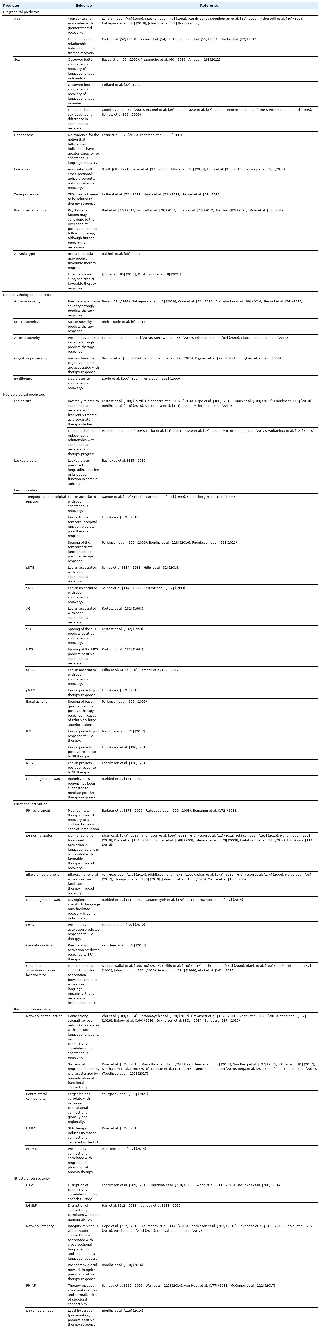
Table 1 presents an overview of the evidence reviewed above. In terms of biographical predictors, age and aphasia type were identified as potential prognostic factors, whereas the relationship between psychosocial factors and eduction, and therapy outcomes remains unclear. Sex, handedness, and time post-onset do not seem to influence treated recovery. Pre-therapy aphasia severity was the most consistently identified neuropsychological predictor, and variable measures of cognitive processing were independently associated with therapy outcomes in several studies. No evidence was found for an effect of nonverbal intelligence on language outcomes. As far as sheer volume goes, most research has been conducted on neurobiological predictors. Factors such as lesion size and location (e.g., damage to the temporoparietal junction) have frequently been associated with extent of recovery, and optimal recovery may rely on functionality of residual language regions within the LH. Similarly, integrity of the functional and structural connectomes, especially in frontal (e.g., Broca’s area) and posterior temporal areas has been found to facilitate successful response to therapy.

Predictors of aphasia recovery

Collectively, although generalizable prognostic factors are scarce, these findings reveal that it is certainly possible to model therapy response based on biographical, neuropsychological, and neurobiological data collected prior to therapy. The failure of previous research to generate clinically applicable predictors does not represent that this endeavor is impossible; rather, as noted above, it is a manifestation of small sample sizes. The same issue is not as prevalent in cross-sectional studies and studies on spontaneous recovery, as these study designs typically include a greater number of participants [188,193,203] and, therefore, achieve greater statistical power. However, therapy studies are time-consuming, expensive, and require more resources. Thus, the challenge of prior studies to address the fundamental questions of who responds to therapy and to what degree likely reflects a shortage of the resources necessary to adequately address them.

Few researchers would argue against the notion that the literature clearly demonstrates the potential to improve prognostication in chronic aphasia. As a testimony to this end, the call for advancement of personalized solutions in aphasia therapy has grown immensely in recent years [15,16,105,223]. We would argue that the first step to meet the increasing demand for PAT will be to emphasize participant recruitment on a grander scale. In fact, such efforts are currently underway. Recent multisite collaborative efforts (e.g., Center for the Study of Aphasia Recovery [C-STAR], Predicting Language Outcome and Recovery After Stroke [PLORAS]) enable participant recruitment on a grander scale and, therefore, offer increased statistical power to counter the heterogeneous characteristics of this population. Further, larger sample sizes will be integral to examine how different predictors (e.g., age and lesion characteristics) support or deter recovery, and to examine how predictors interact with other therapy parameters such as dose, intensity, and therapy type. These collaborative initiatives hold tremendous promise to address some of the persistent issues described above.

The potential implications of identifying robust and generalizable predictors of therapy response in aphasia are substantial. In terms of clinical practice, improved understanding of therapy response will enable clinicians to personalize aphasia therapy more efficiently and, by extension, to enhance therapy outcomes. As for research perspectives, understanding therapy response at the individual level is fundamental for future efforts to identify what type of therapy works for whom, e.g., who is likely to benefit from pharmacotherapy, non-invasive brain stimulation, and neurofeedback training; which individuals benefit from intensive or distributed therapy; and, equally importantly, what should the focus of treatment be for individuals unlikely to respond to conventional impairment-based SLT. Thus, we believe these clear advantages of pursuing larger-scale, scientifically rigorous study designs outweigh any potential disadvantages associated with greater demand for tangible resources. Ultimately, and most importantly, these research efforts will pay dividends to PWA through substantially improved quality of life.

Conclusions

Efforts to identify robust predictors of therapy response in chronic aphasia have, as of yet, failed to generate clinically applicable findings. As a result, clinicians cannot reliably determine who is likely to respond to impairment-based therapy and to what degree, with reference to standardized clinical guidelines. Our review reveals that while several biographical, neuropsychological, and neurobiological predictors have consistently been reported, most findings cannot be generalized to the population of PWA due to use of small sample sizes. Further, inconsistent and mixed findings are a prevalent problem, likely for the same reason. Identifying reliable predictors of therapy response is a necessary prerequisite for the development of personalized therapy solutions in aphasia, i.e., therapy tailored to maximize each individual’s language recovery. Future research should aim to recruit a greater number of participants to actively facilitate the advancement of clinical management of aphasia.

Notes

Disclosure

The authors have no financial conflicts of interest.

References

1. Mesulam MM. Aphasia, sudden and progressive. In : Whitaker HA, ed. Concise Encyclopedia of Brain and Language Oxford, UK: Elsevier Ltd; 2010. p. 49–53.
2. Watila MM, Balarabe SA. Factors predicting post-stroke aphasia recovery. J Neurol Sci 2015;352:12–18.
3. Code C, Papathanasiou I, Rubio-Bruno S, Cabana Mde L, Villanueva MM, Haaland-Johansen L, et al. International patterns of the public awareness of aphasia. Int J Lang Commun Disord 2016;51:276–284.
4. Flowers HL, Skoretz SA, Silver FL, Rochon E, Fang J, Flamand-Roze C, et al. Poststroke aphasia frequency, recovery, and outcomes: a systematic review and meta-analysis. Arch Phys Med Rehabil 2016;97:2188–2201.
5. Brady MC, Kelly H, Godwin J, Enderby P, Campbell P. Speech and language therapy for aphasia following stroke. Cochrane Database Syst Rev 2016;2016:CD000425.
6. Breitenstein C, Grewe T, Flöel A, Ziegler W, Springer L, Martus P, et al. Intensive speech and language therapy in patients with chronic aphasia after stroke: a randomised, open-label, blinded-endpoint, controlled trial in a health-care setting. Lancet 2017;389:1528–1538.
7. Fama ME, Turkeltaub PE. Treatment of poststroke aphasia: current practice and new directions. Semin Neurol 2014;34:504–513.
8. Kristinsson S, Basilakos A, Elm J, Spell LA, Bonilha L, Rorden C, et al. Individualized response to semantic versus phonological aphasia therapies in stroke. Brain Commun 2021;3:fcab174.
9. Menahemi-Falkov M, Breitenstein C, Pierce JE, Hill AJ, O’Halloran R, Rose ML. A systematic review of maintenance following intensive therapy programs in chronic post-stroke aphasia: importance of individual response analysis. Disabil Rehabil 2021;Aug. 12. [Epub]. https://doi.org/10.1080/09638288.2021.1955303.
10. Price CJ. The anatomy of language: a review of 100 fMRI studies published in 2009. Ann N Y Acad Sci 2010;1191:62–88.
11. Fridriksson J, Richardson JD, Fillmore P, Cai B. Left hemisphere plasticity and aphasia recovery. Neuroimage 2012;60:854–863.
12. Lambon Ralph MA, Snell C, Fillingham JK, Conroy P, Sage K. Predicting the outcome of anomia therapy for people with aphasia post CVA: both language and cognitive status are key predictors. Neuropsychol Rehabil 2010;20:289–305.
13. Schevenels K, Price CJ, Zink I, De Smedt B, Vandermosten M. A review on treatment-related brain changes in aphasia. Neurobiol Lang 2020;1:402–433.
14. Cheng BB, Worrall LE, Copland DA, Wallace SJ. Prognostication in post-stroke aphasia: how do speech pathologists formulate and deliver information about recovery? Int J Lang Commun Disord 2020;55:520–536.
15. Berube S, Hillis AE. Advances and innovations in aphasia treatment trials. Stroke 2019;50:2977–2984.
16. Doogan C, Dignam J, Copland D, Leff A. Aphasia recovery: when, how and who to treat? Curr Neurol Neurosci Rep 2018;18:90.
17. Fridriksson J, Hillis AE. Current approaches to the treatment of post-stroke aphasia. J Stroke 2021;23:183–201.
18. Lorca-Puls DL, Gajardo-Vidal A, White J, Seghier ML, Leff AP, Green DW, et al. The impact of sample size on the reproducibility of voxel-based lesion-deficit mappings. Neuropsychologia 2018;115:101–111.
19. Wilson SM, Schneck SM. Neuroplasticity in post-stroke aphasia: a systematic review and meta-analysis of functional imaging studies of reorganization of language processing. Neurobiol Lang (Camb) 2021;2:22–82.
20. Simmons-Mackie N. Aphasia in North America Moorestown, NJ: Aphasia Access; 2018.
21. Boehme AK, Martin-Schild S, Marshall RS, Lazar RM. Effect of aphasia on acute stroke outcomes. Neurology 2016;87:2348–2354.
22. Godecke E, Armstrong E, Rai T, Ciccone N, Rose ML, Middleton S, et al. A randomized control trial of intensive aphasia therapy after acute stroke: the Very Early Rehabilitation for SpEech (VERSE) study. Int J Stroke 2021;16:556–572.
23. Laska AC, Kahan T, Hellblom A, Murray V, von Arbin M. A randomized controlled trial on very early speech and language therapy in acute stroke patients with aphasia. Cerebrovasc Dis Extra 2011;1:66–74.
24. Nouwens F, de Lau LM, Visch-Brink EG, van de Sandt-Koenderman WM, Lingsma HF, Goosen S, et al. Efficacy of early cognitive-linguistic treatment for aphasia due to stroke: a randomised controlled trial (Rotterdam Aphasia Therapy Study-3). Eur Stroke J 2017;2:126–136.
25. Crosson B, Rodriguez AD, Copland D, Fridriksson J, Krishnamurthy LC, Meinzer M, et al. Neuroplasticity and aphasia treatments: new approaches for an old problem. J Neurol Neurosurg Psychiatry 2019;90:1147–1155.
26. Shafto MA, Tyler LK. Language in the aging brain: the network dynamics of cognitive decline and preservation. Science 2014;346:583–587.
27. Vara H, Muñoz-Cuevas J, Colino A. Age-dependent alterations of long-term synaptic plasticity in thyroid-deficient rats. Hippocampus 2003;13:816–825.
28. deToledo-Morrell L, Geinisman Y, Morrell F. Age-dependent alterations in hippocampal synaptic plasticity: relation to memory disorders. Neurobiol Aging 1988;9:581–590.
29. REhabilitation and recovery of peopLE with Aphasia after StrokE (RELEASE) Collaborators. Predictors of poststroke aphasia recovery: a systematic review-informed individual participant data meta-analysis. Stroke 2021;52:1778–1787.
30. El Hachioui H, Lingsma HF, van de Sandt-Koenderman MW, Dippel DW, Koudstaal PJ, Visch-Brink EG. Long-term prognosis of aphasia after stroke. J Neurol Neurosurg Psychiatry 2013;84:310–315.
31. Hillis AE, Beh YY, Sebastian R, Breining B, Tippett DC, Wright A, et al. Predicting recovery in acute poststroke aphasia. Ann Neurol 2018;83:612–622.
32. Holland AL, Greenhouse JB, Fromm D, Swindell CS. Predictors of language restitution following stroke: a multivariate analysis. J Speech Hear Res 1989;32:232–238.
33. Kertesz A, McCabe P. Recovery patterns and prognosis in aphasia. Brain 1977;100 Pt 1:1–18.
34. Laska AC, Hellblom A, Murray V, Kahan T, Von Arbin M. Aphasia in acute stroke and relation to outcome. J Intern Med 2001;249:413–422.
35. Marshall RC, Phillips DS. Prognosis for improved verbal communication in aphasic stroke patients. Arch Phys Med Rehabil 1983;64:597–600.
36. Inatomi Y, Yonehara T, Omiya S, Hashimoto Y, Hirano T, Uchino M. Aphasia during the acute phase in ischemic stroke. Cerebrovasc Dis 2008;25:316–323.
37. Lazar RM, Antoniello D. Variability in recovery from aphasia. Curr Neurol Neurosci Rep 2008;8:497–502.
38. Lendrem W, Lincoln NB. Spontaneous recovery of language in patients with aphasia between 4 and 34 weeks after stroke. J Neurol Neurosurg Psychiatry 1985;48:743–748.
39. Pedersen PM, Jørgensen HS, Nakayama H, Raaschou HO, Olsen TS. Aphasia in acute stroke: incidence, determinants, and recovery. Ann Neurol 1995;38:659–666.
40. Pedersen PM, Vinter K, Olsen TS. Aphasia after stroke: type, severity and prognosis: the Copenhagen aphasia study. Cerebrovasc Dis 2004;17:35–43.
41. Engelter ST, Gostynski M, Papa S, Frei M, Born C, Ajdacic-Gross V, et al. Epidemiology of aphasia attributable to first ischemic stroke: incidence, severity, fluency, etiology, and thrombolysis. Stroke 2006;37:1379–1384.
42. Miceli G, Caltagirone C, Gainotti G, Masullo C, Silveri MC, Villa G. Influence of age, sex, literacy and pathologic lesion on incidence, severity and type of aphasia. Acta Neurol Scand 1981;64:370–382.
43. De Renzi E, Faglioni P, Ferrari P. The influence of sex and age on the incidence and type of aphasia. Cortex 1980;16:627–630.
44. Ferro JM, Madureira S. Aphasia type, age and cerebral infarct localisation. J Neurol 1997;244:505–509.
45. Smith A. Ambiguities in concepts and studies of “brain damage” and “organicity”. J Nerv Ment Dis 1962;135:311–326.
46. Lendrem W, McGuirk E, Lincoln NB. Factors affecting language recovery in aphasic stroke patients receiving speech therapy. J Neurol Neurosurg Psychiatry 1988;51:1103–1104.
47. Marshall RC, Tompkins CA, Phillips DS. Improvement in treated aphasia: examination of selected prognostic factors. Folia Phoniatr (Basel) 1982;34:305–315.
48. Nakagawa Y, Sano Y, Funayama M, Kato M. Prognostic factors for long-term improvement from stroke-related aphasia with adequate linguistic rehabilitation. Neurol Sci 2019;40:2141–2146.
49. Pickersgill MJ, Lincoln NB. Prognostic indicators and the pattern of recovery of communication in aphasic stroke patients. J Neurol Neurosurg Psychiatry 1983;46:130–139.
50. van de Sandt-Koenderman WM, van Harskamp F, Duivenvoorden HJ, Remerie SC, van der Voort-Klees YA, Wielaert SM, et al. MAAS (Multi-axial Aphasia System): realistic goal setting in aphasia rehabilitation. Int J Rehabil Res 2008;31:314–320.
51. Johnson LP, Duffy C, Basilakos A, Rorden C, Bonilha L, Fridriksson J. Age and initial severity as predictors of treatment outcomes in chronic post-stroke aphasia. Am J Speech Lang Pathol Forthcoming 2022.
52. Code C, Torney A, Gildea-Howardine E, Willmes K. Outcome of a one-month therapy intensive for chronic aphasia: variable individual responses. Semin Speech Lang 2010;31:21–33.
53. Nardo D, Holland R, Leff AP, Price CJ, Crinion JT. Less is more: neural mechanisms underlying anomia treatment in chronic aphasic patients. Brain 2017;140:3039–3054.
54. Persad C, Wozniak L, Kostopoulos E. Retrospective analysis of outcomes from two intensive comprehensive aphasia programs. Top Stroke Rehabil 2013;20:388–397.
55. Seniów J, Litwin M, Leśniak M. The relationship between non-linguistic cognitive deficits and language recovery in patients with aphasia. J Neurol Sci 2009;283:91–94.
56. Ellis C, Urban S. Age and aphasia: a review of presence, type, recovery and clinical outcomes. Top Stroke Rehabil 2016;23:430–439.
57. Wallentin M. Sex differences in post-stroke aphasia rates are caused by age: a meta-analysis and database query. PLoS One 2018;13:e0209571.
58. Shaywitz BA, Shaywitz SE, Pugh KR, Constable RT, Skudlarski P, Fulbright RK, et al. Sex differences in the functional organization of the brain for language. Nature 1995;373:607–609.
59. Basso A. Prognostic factors in aphasia. Aphasiology 1992;6:337–348.
60. Pizzamiglio L, Mammucari A, Razzano C. Evidence for sex differences in brain organization in recovery in aphasia. Brain Lang 1985;25:213–223.
61. Godefroy O, Dubois C, Debachy B, Leclerc M, Kreisler A, ; Lille Stroke Program. Vascular aphasias: main characteristics of patients hospitalized in acute stroke units. Stroke 2002;33:702–705.
62. Szaflarski JP, Rajagopal A, Altaye M, Byars AW, Jacola L, Schmithorst VJ, et al. Left-handedness and language lateralization in children. Brain Res 2012;1433:85–97.
63. Szaflarski JP, Binder JR, Possing ET, McKiernan KA, Ward BD, Hammeke TA. Language lateralization in left-handed and ambidextrous people: fMRI data. Neurology 2002;59:238–244.
64. Szaflarski JP, Holland SK, Schmithorst VJ, Byars AW. fMRI study of language lateralization in children and adults. Hum Brain Mapp 2006;27:202–212.
65. Hillis AE, Tippett DC. Stroke recovery: surprising influences and residual consequences. Adv Med 2014;2014:378263.
66. Lazar RM, Minzer B, Antoniello D, Festa JR, Krakauer JW, Marshall RS. Improvement in aphasia scores after stroke is well predicted by initial severity. Stroke 2010;41:1485–1488.
67. Ramsey LE, Siegel JS, Lang CE, Strube M, Shulman GL, Corbetta M. Behavioural clusters and predictors of performance during recovery from stroke. Nat Hum Behav 2017;1:0038.
68. Smith A. Objective indices of severity of chronic aphasia in stroke patients. J Speech Hear Disord 1971;36:167–207.
69. Connor LT, Obler LK, Tocco M, Fitzpatrick PM, Albert ML. Effect of socioeconomic status on aphasia severity and recovery. Brain Lang 2001;78:254–257.
70. Plowman E, Hentz B, Ellis C Jr. Post-stroke aphasia prognosis: a review of patient-related and stroke-related factors. J Eval Clin Pract 2012;18:689–694.
71. Kertesz A. Recovery and treatment. Heilman KM, Valenstein E. Clinical Neuropsychology 3rd edth ed. Oxford, UK: Oxford University Press; 1993. p. 647–674.
72. Holland A, Fromm D, Forbes M, MacWhinney B. Long-term recovery in stroke accompanied by aphasia: a reconsideration. Aphasiology 2017;31:152–165.
73. Moss A, Nicholas M. Language rehabilitation in chronic aphasia and time postonset: a review of single-subject data. Stroke 2006;37:3043–3051.
74. Hilari K, Needle JJ, Harrison KL. What are the important factors in health-related quality of life for people with aphasia?: a systematic review. Arch Phys Med Rehabil 2012;93(1 Suppl):S86–S95.
75. Hilari K, Northcott S. Social support in people with chronic aphasia. Aphasiology 2006;20:17–36.
76. Worrall LE, Hudson K, Khan A, Ryan B, Simmons-Mackie N. Determinants of living well with aphasia in the first year poststroke: a prospective cohort study. Arch Phys Med Rehabil 2017;98:235–240.
77. Biel M, Nitta L, Jackson C. Understanding motivation in aphasia rehabilitation. In : Coppens P, Patterson JL, eds. Aphasia Rehabilitation: Clinical Challenges Burlington, MA: Jones & Bartlett Learning; 2017. p. 393–436.
78. Chapey R, Duchan JF, Elman RJ, Garcia LJ, Kagan A, Lyon JG, et al. Life participation approach to aphasia: a statement of values for the future. ASHA Lead 2000;5:4–6.
79. Attard MC, Lanyon L, Togher L, Rose ML. Consumer perspectives on community aphasia groups: a narrative literature review in the context of psychological well-being. Aphasiology 2015;29:983–1019.
80. Attard MC, Loupis Y, Togher L, Rose ML. Experiences of people with severe aphasia and spouses attending an Interdisciplinary Community Aphasia Group. Disabil Rehabil 2020;42:1382–1396.
81. Lanyon L, Worrall L, Rose M. Combating social isolation for people with severe chronic aphasia through community aphasia groups: consumer views on getting it right and wrong. Aphasiology 2018;32:493–517.
82. Berthier ML. Ten key reasons for continuing research on pharmacotherapy for post-stroke aphasia. Aphasiology 2021;35:824–858.
83. Mohr B, Stahl B, Berthier ML, Pulvermüller F. Intensive communicative therapy reduces symptoms of depression in chronic nonfluent aphasia. Neurorehabil Neural Repair 2017;31:1053–1062.
84. Thomas SA, Walker MF, Macniven JA, Haworth H, Lincoln NB. Communication and Low Mood (CALM): a randomized controlled trial of behavioural therapy for stroke patients with aphasia. Clin Rehabil 2013;27:398–408.
85. Bakheit AM, Shaw S, Barrett L, Wood J, Carrington S, Griffiths S, et al. A prospective, randomized, parallel group, controlled study of the effect of intensity of speech and language therapy on early recovery from poststroke aphasia. Clin Rehabil 2007;21:885–894.
86. Jung IY, Lim JY, Kang EK, Sohn HM, Paik NJ. The factors associated with good responses to speech therapy combined with transcranial direct current stimulation in post-stroke aphasic patients. Ann Rehabil Med 2011;35:460–469.
87. Wade DT, Hewer RL, David RM, Enderby PM. Aphasia after stroke: natural history and associated deficits. J Neurol Neurosurg Psychiatry 1986;49:11–16.
88. Efstratiadou EA, Papathanasiou I, Holland R, Archonti A, Hilari K. A systematic review of semantic feature analysis therapy studies for aphasia. J Speech Lang Hear Res 2018;61:1261–1278.
89. Wisenburn B, Mahoney K. A meta-analysis of word-finding treatments for aphasia. Aphasiology 2009;23:1338–1352.
90. Robey RR. A meta-analysis of clinical outcomes in the treatment of aphasia. J Speech Lang Hear Res 1998;41:172–187.
91. Beeson PM, Bayles KA, Rubens AB, Kaszniak AW. Memory impairment and executive control in individuals with stroke-induced aphasia. Brain Lang 1993;45:253–275.
92. Burgio F, Basso A. Memory and aphasia. Neuropsychologia 1997;35:759–766.
93. Helm-Estabrooks N. Cognition and aphasia: a discussion and a study. J Commun Disord 2002;35:171–186.
94. Martin N, Gupta P. Exploring the relationship between word processing and verbal short-term memory: evidence from associations and dissociations. Cogn Neuropsychol 2004;21:213–228.
95. Murray L, Salis C, Martin N, Dralle J. The use of standardised short-term and working memory tests in aphasia research: a systematic review. Neuropsychol Rehabil 2018;28:309–351.
96. Marinelli CV, Spaccavento S, Craca A, Marangolo P, Angelelli P. Different cognitive profiles of patients with severe aphasia. Behav Neurol 2017;2017:3875954.
97. Dignam J, Copland D, O’Brien K, Burfein P, Khan A, Rodriguez AD. Influence of cognitive ability on therapy outcomes for anomia in adults with chronic poststroke aphasia. J Speech Lang Hear Res 2017;60:406–421.
98. Fillingham JK, Sage K, Lambon Ralph MA. The treatment of anomia using errorless learning. Neuropsychol Rehabil 2006;16:129–154.
99. Kertesz A, McCabe P. Intelligence and aphasia: performance of aphasics on Raven’s coloured progressive matrices (RCPM). Brain Lang 1975;2:387–395.
100. David RM, Skilbeck CE. Raven IQ and language recovery following stroke. J Clin Neuropsychol 1984;6:302–308.
101. Ferro JM, Mariano G, Madureira S. Recovery from aphasia and neglect. Cerebrovasc Dis 1999;9 Suppl 5:6–22.
102. Broca P. Remarques sur le siege de la faculte du langage articule, suivies d’une observation d’aphemie (perte de la parole). Bullet Soci Anatom Paris 1861;6:330–357.
103. Broca P. Sur le siege de la faculte du langage articule. Bull Mem Soc Anthropol Paris 1865;6:377–393.
104. Wernicke C. Der Aphasische Symptomencomplex Breslau, PL: Cohn and Weigert; 1874.
105. Kiran S, Thompson CK. Neuroplasticity of language networks in aphasia: advances, updates, and future challenges. Front Neurol 2019;10:295.
106. Hope TM, Seghier ML, Leff AP, Price CJ. Predicting outcome and recovery after stroke with lesions extracted from MRI images. Neuroimage Clin 2013;2:424–433.
107. Goldenberg G, Spatt J. Influence of size and site of cerebral lesions on spontaneous recovery of aphasia and on success of language therapy. Brain Lang 1994;47:684–698.
108. Kertesz A, Harlock W, Coates R. Computer tomographic localization, lesion size, and prognosis in aphasia and nonverbal impairment. Brain Lang 1979;8:34–50.
109. Maas MB, Lev MH, Ay H, Singhal AB, Greer DM, Smith WS, et al. The prognosis for aphasia in stroke. J Stroke Cerebrovasc Dis 2012;21:350–357.
110. Mazzoni M, Vista M, Pardossi L, Avila L, Bianchi F, Moretti P. Spontaneous evolution of aphasia after ischaemic stroke. Aphasiology 1992;6:387–396.
111. Naeser MA, Palumbo CL. Neuroimaging and language recovery in stroke. J Clin Neurophysiol 1994;11:150–174.
112. Naeser MA, Helm-Estabrooks N, Haas G, Auerbach S, Srinivasan M. Relationship between lesion extent in ‘Wernicke’s area’ on computed tomographic scan and predicting recovery of comprehension in Wernicke’s aphasia. Arch Neurol 1987;44:73–82.
113. Naeser MA, Palumbo CL, Helm-Estabrooks N, Stiassny-Eder D, Albert ML. Severe nonfluency in aphasia: role of the medial subcallosal fasciculus and other white matter pathways in recovery of spontaneous speech. Brain 1989;112(Pt 1):1–38.
114. Selnes OA, Knopman DS, Niccum N, Rubens AB, Larson D. Computed tomographic scan correlates of auditory comprehension deficits in aphasia: a prospective recovery study. Ann Neurol 1983;13:558–566.
115. Basilakos A, Stark BC, Johnson L, Rorden C, Yourganov G, Bonilha L, et al. Leukoaraiosis is associated with a decline in language abilities in chronic aphasia. Neurorehabil Neural Repair 2019;33:718–729.
116. Kristinsson S, Zhang W, Rorden C, Newman-Norlund R, Basilakos A, Bonilha L, et al. Machine learning-based multimodal prediction of language outcomes in chronic aphasia. Hum Brain Mapp 2021;42:1682–1698.
117. Yourganov G, Fridriksson J, Rorden C, Gleichgerrcht E, Bonilha L. Multivariate connectome-based symptom mapping in post-stroke patients: networks supporting language and speech. J Neurosci 2016;36:6668–6679.
118. Bonilha L, Gleichgerrcht E, Nesland T, Rorden C, Fridriksson J. Success of anomia treatment in aphasia is associated with preserved architecture of global and left temporal lobe structural networks. Neurorehabil Neural Repair 2016;30:266–279.
119. Fridriksson J. Preservation and modulation of specific left hemisphere regions is vital for treated recovery from anomia in stroke. J Neurosci 2010;30:11558–11564.
120. Meier EL, Johnson JP, Pan Y, Kiran S. The utility of lesion classification in predicting language and treatment outcomes in chronic stroke-induced aphasia. Brain Imaging Behav 2019;13:1510–1525.
121. Varkanitsa M, Peñaloza C, Charidimou A, Caplan D, Kiran S. White matter hyperintensities predict response to language treatment in poststroke aphasia. Neurorehabil Neural Repair 2020;34:945–953.
122. Marcotte K, Adrover-Roig D, Damien B, de Préaumont M, Généreux S, Hubert M, et al. Therapy-induced neuroplasticity in chronic aphasia. Neuropsychologia 2012;50:1776–1786.
123. Payabvash S, Kamalian S, Fung S, Wang Y, Passanese J, Kamalian S, et al. Predicting language improvement in acute stroke patients presenting with aphasia: a multivariate logistic model using location-weighted atlas-based analysis of admission CT perfusion scans. AJNR Am J Neuroradiol 2010;31:1661–1668.
124. Thye M, Mirman D. Relative contributions of lesion location and lesion size to predictions of varied language deficits in post-stroke aphasia. Neuroimage Clin 2018;20:1129–1138.
125. Parkinson BR, Raymer A, Chang YL, Fitzgerald DB, Crosson B. Lesion characteristics related to treatment improvement in object and action naming for patients with chronic aphasia. Brain Lang 2009;110:61–70.
126. Stockert A, Wawrzyniak M, Klingbeil J, Wrede K, Kümmerer D, Hartwigsen G, et al. Dynamics of language reorganization after left temporo-parietal and frontal stroke. Brain 2020;143:844–861.
127. Kristinsson S, Thors H, Yourganov G, Magnusdottir S, Hjaltason H, Stark BC, et al. Brain damage associated with impaired sentence processing in acute aphasia. J Cogn Neurosci 2020;32:256–271.
128. Matchin W, Hickok G. The cortical organization of syntax. Cereb Cortex 2020;30:1481–1498.
129. van den Heuvel MP, Sporns O. Network hubs in the human brain. Trends Cogn Sci 2013;17:683–696.
130. Alexander MP, Naeser MA, Palumbo C. Broca’s area aphasias: aphasia after lesions including the frontal operculum. Neurology 1990;40:353–362.
131. Hanlon RE, Lux WE, Dromerick AW. Global aphasia without hemiparesis: language profiles and lesion distribution. J Neurol Neurosurg Psychiatry 1999;66:365–369.
132. Kertesz A, Lau WK, Polk M. The structural determinants of recovery in Wernicke’s aphasia. Brain Lang 1993;44:153–164.
133. Naeser MA, Gaddie A, Palumbo CL, Stiassny-Eder D. Late recovery of auditory comprehension in global aphasia: improved recovery observed with subcortical temporal isthmus lesion vs Wernicke’s cortical area lesion. Arch Neurol 1990;47:425–432.
134. Benghanem S, Rosso C, Arbizu C, Moulton E, Dormont D, Leger A, et al. Aphasia outcome: the interactions between initial severity, lesion size and location. J Neurol 2019;266:1303–1309.
135. Kang EK, Sohn HM, Han MK, Kim W, Han TR, Paik NJ. Severity of post-stroke aphasia according to aphasia type and lesion location in Koreans. J Korean Med Sci 2010;25:123–127.
136. Fridriksson J, Basilakos A, Hickok G, Bonilha L, Rorden C. Speech entrainment compensates for Broca’s area damage. Cortex 2015;69:68–75.
137. Brownsett SL, Warren JE, Geranmayeh F, Woodhead Z, Leech R, Wise RJ. Cognitive control and its impact on recovery from aphasic stroke. Brain 2014;137(Pt 1):242–254.
138. Sul B, Lee KB, Hong BY, Kim JS, Kim J, Hwang WS, et al. Association of lesion location with long-term recovery in poststroke aphasia and language deficits. Front Neurol 2019;10:776.
139. Saur D, Lange R, Baumgaertner A, Schraknepper V, Willmes K, Rijntjes M, et al. Dynamics of language reorganization after stroke. Brain 2006;129(Pt 6):1371–1384.
140. Crinion JT, Leff AP. Recovery and treatment of aphasia after stroke: functional imaging studies. Curr Opin Neurol 2007;20:667–673.
141. Crinion JT, Leff AP. Using functional imaging to understand therapeutic effects in poststroke aphasia. Curr Opin Neurol 2015;28:330–337.
142. Crosson B, McGregor K, Gopinath KS, Conway TW, Benjamin M, Chang YL, et al. Functional MRI of language in aphasia: a review of the literature and the methodological challenges. Neuropsychol Rev 2007;17:157–177.
143. Meinzer M, Harnish S, Conway T, Crosson B. Recent developments in functional and structural imaging of aphasia recovery after stroke. Aphasiology 2011;25:271–290.
144. Thompson CK, den Ouden DB. Neuroimaging and recovery of language in aphasia. Curr Neurol Neurosci Rep 2008;8:475–483.
145. Zahn R, Schwarz M, Huber W. Functional activation studies of word processing in the recovery from aphasia. J Physiol Paris 2006;99:370–385.
146. Heiss WD, Thiel A. A proposed regional hierarchy in recovery of post-stroke aphasia. Brain Lang 2006;98:118–123.
147. Fridriksson J, Bonilha L, Baker JM, Moser D, Rorden C. Activity in preserved left hemisphere regions predicts anomia severity in aphasia. Cereb Cortex 2010;20:1013–1019.
148. Griffis JC, Nenert R, Allendorfer JB, Vannest J, Holland S, Dietz A, et al. The canonical semantic network supports residual language function in chronic post-stroke aphasia. Hum Brain Mapp 2017;38:1636–1658.
149. Karbe H, Thiel A, Weber-Luxenburger G, Herholz K, Kessler J, Heiss WD. Brain plasticity in poststroke aphasia: what is the contribution of the right hemisphere? Brain Lang 1998;64:215–230.
150. Nenert R, Allendorfer JB, Martin AM, Banks C, Vannest J, Holland SK, et al. Longitudinal fMRI study of language recovery after a left hemispheric ischemic stroke. Restor Neurol Neurosci 2018;36:359–385.
151. Szaflarski JP, Allendorfer JB, Banks C, Vannest J, Holland SK. Recovered vs. not-recovered from post-stroke aphasia: the contributions from the dominant and non-dominant hemispheres. Restor Neurol Neurosci 2013;31:347–360.
152. Tyler LK, Wright P, Randall B, Marslen-Wilson WD, Stamatakis EA. Reorganization of syntactic processing following left-hemisphere brain damage: does right-hemisphere activity preserve function? Brain 2010;133:3396–3408.
153. Warburton E, Price CJ, Swinburn K, Wise RJ. Mechanisms of recovery from aphasia: evidence from positron emission tomography studies. J Neurol Neurosurg Psychiatry 1999;66:155–161.
154. Winhuisen L, Thiel A, Schumacher B, Kessler J, Rudolf J, Haupt WF, et al. The right inferior frontal gyrus and poststroke aphasia: a follow-up investigation. Stroke 2007;38:1286–1292.
155. Cappa SF, Perani D, Grassi F, Bressi S, Alberoni M, Franceschi M, et al. A PET follow-up study of recovery after stroke in acute aphasics. Brain Lang 1997;56:55–67.
156. Kinsbourne M. The minor cerebral hemisphere as a source of aphasic speech. Arch Neurol 1971;25:302–306.
157. Leff A, Crinion J, Scott S, Turkheimer F, Howard D, Wise R. A physiological change in the homotopic cortex following left posterior temporal lobe infarction. Ann Neurol 2002;51:553–558.
158. Ohyama M, Senda M, Kitamura S, Ishii K, Mishina M, Terashi A. Role of the nondominant hemisphere and undamaged area during word repetition in poststroke aphasics: a PET activation study. Stroke 1996;27:897–903.
159. Raboyeau G, De Boissezon X, Marie N, Balduyck S, Puel M, Bézy C, et al. Right hemisphere activation in recovery from aphasia: lesion effect or function recruitment? Neurology 2008;70:290–298.
160. Weiller C, Isensee C, Rijntjes M, Huber W, Müller S, Bier D, et al. Recovery from Wernicke’s aphasia: a positron emission tomographic study. Ann Neurol 1995;37:723–732.
161. Abel S, Weiller C, Huber W, Willmes K, Specht K. Therapy-induced brain reorganization patterns in aphasia. Brain 2015;138(Pt 4):1097–1112.
162. Musso M, Weiller C, Kiebel S, Müller SP, Bülau P, Rijntjes M. Training-induced brain plasticity in aphasia. Brain 1999;122(Pt 9):1781–1790.
163. Price CJ, Warburton EA, Moore CJ, Frackowiak RS, Friston KJ. Dynamic diaschisis: anatomically remote and context-sensitive human brain lesions. J Cogn Neurosci 2001;13:419–429.
164. Dietz A, Vannest J, Maloney T, Altaye M, Holland S, Szaflarski JP. The feasibility of improving discourse in people with aphasia through AAC: clinical and functional MRI correlates. Aphasiology 2018;32:693–719.
165. Hallam GP, Thompson HE, Hymers M, Millman RE, Rodd JM, Lambon Ralph MA, et al. Task-based and resting-state fMRI reveal compensatory network changes following damage to left inferior frontal gyrus. Cortex 2018;99:150–165.
166. Johnson JP, Meier EL, Pan Y, Kiran S. Pre-treatment graph measures of a functional semantic network are associated with naming therapy outcomes in chronic aphasia. Brain Lang 2020;207:104809.
167. Kiran S, Meier EL, Johnson JP. Neuroplasticity in aphasia: a proposed framework of language recovery. J Speech Lang Hear Res 2019;62:3973–3985.
168. Richter M, Miltner WH, Straube T. Association between therapy outcome and right-hemispheric activation in chronic aphasia. Brain 2008;131(Pt 5):1391–1401.
169. Thompson CK, Riley EA, den Ouden DB, Meltzer-Asscher A, Lukic S. Training verb argument structure production in agrammatic aphasia: behavioral and neural recovery patterns. Cortex 2013;49:2358–2376.
170. Meinzer M, Breitenstein C. Functional imaging studies of treatment‐induced recovery in chronic aphasia. Aphasiology 2008;22:1251–1268.
171. Barbieri E, Mack J, Chiappetta B, Europa E, Thompson CK. Recovery of offline and online sentence processing in aphasia: language and domain-general network neuroplasticity. Cortex 2019;120:394–418.
172. Benjamin EJ, Virani SS, Callaway CW, Chamberlain AM, Chang AR, Cheng S, et al. Heart disease and stroke statistics-2018 update: a report from the American Heart Association. Circulation 2018;137:e67–e492.
173. Fridriksson J, Moser D, Bonilha L, Morrow-Odom KL, Shaw H, Fridriksson A, et al. Neural correlates of phonological and semantic-based anomia treatment in aphasia. Neuropsychologia 2007;45:1812–1822.
174. Fridriksson J, Nettles C, Davis M, Morrow L, Montgomery A. Functional communication and executive function in aphasia. Clin Linguist Phon 2006;20:401–410.
175. Kiran S, Meier EL, Kapse KJ, Glynn PA. Changes in task-based effective connectivity in language networks following rehabilitation in post-stroke patients with aphasia. Front Hum Neurosci 2015;9:316.
176. Thompson CK, Bonakdarpour B, Fix SF. Neural mechanisms of verb argument structure processing in agrammatic aphasic and healthy age-matched listeners. J Cogn Neurosci 2010;22:1993–2011.
177. van Hees S, McMahon K, Angwin A, de Zubicaray G, Read S, Copland DA. A functional MRI study of the relationship between naming treatment outcomes and resting state functional connectivity in post-stroke aphasia. Hum Brain Mapp 2014;35:3919–3931.
178. Geranmayeh F, Chau TW, Wise RJ, Leech R, Hampshire A. Domain-general subregions of the medial prefrontal cortex contribute to recovery of language after stroke. Brain 2017;140:1947–1958.
179. Abel S, Weiller C, Huber W, Willmes K. Neural underpinnings for model-oriented therapy of aphasic word production. Neuropsychologia 2014;57:154–165.
180. Menke R, Meinzer M, Kugel H, Deppe M, Baumgärtner A, Schiffbauer H, et al. Imaging short- and long-term training success in chronic aphasia. BMC Neurosci 2009;10:118.
181. Leonard C, Rochon E, Laird L. Treating naming impairments in aphasia: findings from a phonological components analysis treatment. Aphasiology 2008;22:923–947.
182. Cappa SF. Neuroimaging of recovery from aphasia. Neuropsychol Rehabil 2000;10:365–376.
183. Blank SC, Scott SK, Murphy K, Warburton E, Wise RJ. Speech production: Wernicke, Broca and beyond. Brain 2002;125(Pt 8):1829–1838.
184. Heiss WD, Kessler J, Thiel A, Ghaemi M, Karbe H. Differential capacity of left and right hemispheric areas for compensation of poststroke aphasia. Ann Neurol 1999;45:430–438.
185. Skipper-Kallal LM, Lacey EH, Xing S, Turkeltaub PE. Right hemisphere remapping of naming functions depends on lesion size and location in poststroke aphasia. Neural Plast 2017;2017:8740353.
186. Skipper-Kallal LM, Lacey EH, Xing S, Turkeltaub PE. Functional activation independently contributes to naming ability and relates to lesion site in post-stroke aphasia. Hum Brain Mapp 2017;38:2051–2066.
187. Sandberg CW. Hypoconnectivity of resting-state networks in persons with aphasia compared with healthy age-matched adults. Front Hum Neurosci 2017;11:91.
188. Siegel JS, Seitzman BA, Ramsey LE, Ortega M, Gordon EM, Dosenbach NU, et al. Re-emergence of modular brain networks in stroke recovery. Cortex 2018;101:44–59.
189. Zhu D, Chang J, Freeman S, Tan Z, Xiao J, Gao Y, et al. Changes of functional connectivity in the left frontoparietal network following aphasic stroke. Front Behav Neurosci 2014;8:167.
190. Balaev V, Petrushevsky A, Martynova O. Changes in functional connectivity of default mode network with auditory and right frontoparietal networks in poststroke aphasia. Brain Connect 2016;6:714–723.
191. Dijkhuizen RM, Zaharchuk G, Otte WM. Assessment and modulation of resting-state neural networks after stroke. Curr Opin Neurol 2014;27:637–643.
192. Yang M, Li J, Yao D, Chen H. Disrupted intrinsic local synchronization in poststroke aphasia. Medicine (Baltimore) 2016;95:e3101.
193. Yourganov G, Stark BC, Fridriksson J, Bonilha L, Rorden C. Effect of stroke on contralateral functional connectivity. Brain Connect 2021;11:543–552.
194. Duncan ES, Small SL. Increased modularity of resting state networks supports improved narrative production in aphasia recovery. Brain Connect 2016;6:524–529.
195. Gili T, Fiori V, De Pasquale G, Sabatini U, Caltagirone C, Marangolo P. Right sensory-motor functional networks subserve action observation therapy in aphasia. Brain Imaging Behav 2017;11:1397–1411.
196. Marcotte K, Perlbarg V, Marrelec G, Benali H, Ansaldo AI. Default-mode network functional connectivity in aphasia: therapy-induced neuroplasticity. Brain Lang 2013;124:45–55.
197. Sandberg CW, Bohland JW, Kiran S. Changes in functional connectivity related to direct training and generalization effects of a word finding treatment in chronic aphasia. Brain Lang 2015;150:103–116.
198. Santhanam P, Duncan ES, Small SL. Therapy-induced plasticity in chronic aphasia is associated with behavioral improvement and time since stroke. Brain Connect 2018;8:179–188.
199. Baliki MN, Babbitt EM, Cherney LR. Brain network topology influences response to intensive comprehensive aphasia treatment. NeuroRehabilitation 2018;43:63–76.
200. Duncan ES, Small SL. Changes in dynamic resting state network connectivity following aphasia therapy. Brain Imaging Behav 2018;12:1141–1149.
201. Iorga M, Higgins J, Caplan D, Zinbarg R, Kiran S, Thompson CK, et al. Predicting language recovery in post-stroke aphasia using behavior and functional MRI. Sci Rep 2021;11:8419.
202. Woodhead ZV, Crinion J, Teki S, Penny W, Price CJ, Leff AP. Auditory training changes temporal lobe connectivity in ‘Wernicke’s aphasia’: a randomised trial. J Neurol Neurosurg Psychiatry 2017;88:586–594.
203. Fridriksson J, den Ouden DB, Hillis AE, Hickok G, Rorden C, Basilakos A, et al. Anatomy of aphasia revisited. Brain 2018;141:848–862.
204. Mesulam MM. Large-scale neurocognitive networks and distributed processing for attention, language, and memory. Ann Neurol 1990;28:597–613.
205. Catani M, Mesulam M. The arcuate fasciculus and the disconnection theme in language and aphasia: history and current state. Cortex 2008;44:953–961.
206. Gleichgerrcht E, Kocher M, Nesland T, Rorden C, Fridriksson J, Bonilha L. Preservation of structural brain network hubs is associated with less severe post-stroke aphasia. Restor Neurol Neurosci 2016;34:19–28.
207. Forkel SJ, Catani M. Lesion mapping in acute stroke aphasia and its implications for recovery. Neuropsychologia 2018;115:88–100.
208. Basilakos A, Fillmore PT, Rorden C, Guo D, Bonilha L, Fridriksson J. Regional white matter damage predicts speech fluency in chronic post-stroke aphasia. Front Hum Neurosci 2014;8:845.
209. Fridriksson J, Guo D, Fillmore P, Holland A, Rorden C. Damage to the anterior arcuate fasciculus predicts non-fluent speech production in aphasia. Brain 2013;136(Pt 11):3451–3460.
210. Marchina S, Zhu LL, Norton A, Zipse L, Wan CY, Schlaug G. Impairment of speech production predicted by lesion load of the left arcuate fasciculus. Stroke 2011;42:2251–2256.
211. Wang J, Marchina S, Norton AC, Wan CY, Schlaug G. Predicting speech fluency and naming abilities in aphasic patients. Front Hum Neurosci 2013;7:831.
212. Han Z, Ma Y, Gong G, He Y, Caramazza A, Bi Y. White matter structural connectivity underlying semantic processing: evidence from brain damaged patients. Brain 2013;136(Pt 10):2952–2965.
213. Ivanova MV, Isaev DY, Dragoy OV, Akinina YS, Petrushevskiy AG, Fedina ON, et al. Diffusion-tensor imaging of major white matter tracts and their role in language processing in aphasia. Cortex 2016;85:165–181.
214. Del Gaizo J, Fridriksson J, Yourganov G, Hillis AE, Hickok G, Misic B, et al. Mapping language networks using the structural and dynamic brain connectomes. eNeuro 2017;4:ENEURO.0204-17.2017.
215. Halai AD, Woollams AM, Lambon Ralph MA. Investigating the effect of changing parameters when building prediction models for post-stroke aphasia. Nat Hum Behav 2020;4:725–735.
216. Hope TMH, Leff AP, Price CJ. Predicting language outcomes after stroke: is structural disconnection a useful predictor? Neuroimage Clin 2018;19:22–29.
217. Hope TM, Seghier ML, Prejawa S, Leff AP, Price CJ. Distinguishing the effect of lesion load from tract disconnection in the arcuate and uncinate fasciculi. Neuroimage 2016;125:1169–1173.
218. Pustina D, Coslett HB, Ungar L, Faseyitan OK, Medaglia JD, Avants B, et al. Enhanced estimations of post-stroke aphasia severity using stacked multimodal predictions. Hum Brain Mapp 2017;38:5603–5615.
219. Zavanone C, Samson Y, Arbizu C, Dupont S, Dormont D, Rosso C. Critical brain regions related to post-stroke aphasia severity identified by early diffusion imaging are not the same when predicting short- and long-term outcome. Brain Lang 2018;186:1–7.
220. Schlaug G, Marchina S, Norton A. Evidence for plasticity in white-matter tracts of patients with chronic Broca’s aphasia undergoing intense intonation-based speech therapy. Ann N Y Acad Sci 2009;1169:385–394.
221. Wan CY, Zheng X, Marchina S, Norton A, Schlaug G. Intensive therapy induces contralateral white matter changes in chronic stroke patients with Broca’s aphasia. Brain Lang 2014;136:1–7.
222. McKinnon ET, Fridriksson J, Glenn GR, Jensen JH, Helpern JA, Basilakos A, et al. Structural plasticity of the ventral stream and aphasia recovery. Ann Neurol 2017;82:147–151.
223. Simic T, Chambers C, Bitan T, Stewart S, Goldberg D, Laird L, et al. Mechanisms underlying anomia treatment outcomes. J Commun Disord 2020;88:106048.

Article information Continued

Table 1.

Predictors of aphasia recovery

Predictor Evidence Reference
Biographical predictors
Age Younger age is associated with greater treated recovery. Lendrem et al. [46] (1988), Marshall et al. [47] (1982), van de Sandt-Koenderman et al. [50] (2008), Pickersgill et al. [49] (1983), Nakagawa et al. [48] (2019), Johnson et al. [51] (forthcoming)
Failed to find a relationship between age and treated recovery. Code et al. [52] (2010), Persad et al. [54] (2013), Seniów et al. [55] (2009), Nardo et al. [53] (2017)
Sex Observed better spontaneous recovery of language function in females. Basso et al. [59] (1992), Pizzamiglio et al. [60] (1985), Ali et al. [29] (2021)
Observed better spontaneous recovery of language function in males. Holland et al. [32] (1989)
Failed to find a sex-dependent difference in spontaneous recovery. Godefroy et al. [61] (2002), Inatomi et al. [36] (2008), Lazar et al. [37] (2008), Lendrem et al. [38] (1985), Pedersen et al. [39] (1995), Seniów et al. [55] (2009)
Handedness No evidence for the notion that left-handed individuals have greater capacity for spontaneous language recovery. Lazar et al. [37] (2008), Pedersen et al. [39] (1995)
Education Associated with cross-sectional aphasia severity; not spontaneous recovery. Smith [68] (1971), Lazar et al. [37] (2008), Hillis et al. [65] (2014), Hillis et al. [31] (2018), Ramsey et al. [67] (2017)
Time post-onset TPS does not seem to be related to therapy response. Holland et al. [72] (2017), Nardo et al. [53] (2017), Persad et al. [54] (2013)
Psychosocial factors Psychosocial factors may contribute to the likelihood of positive outcomes following therapy, although further research is necessary. Biel et al. [77] (2017), Worrall et al. [76] (2017), Hilari et al. [74] (2012), Berthier [82] (2021), Mohr et al. [83] (2017)
Aphasia type Broca’s aphasia may predict favorable therapy response. Bakheit et al. [85] (2007)
Fluent aphasia subtypes predict favorable therapy response. Jung et al. [86] (2011), Kristinsson et al. [8] (2021)
Neuropsychological predictors
Aphasia severity Pre-therapy aphasia severity strongly predicts therapy response. Basso [59] (1992), Nakagawa et al. [48] (2019), Code et al. [52] (2010), Efstratiadou et al. [88] (2018), Persad et al. [54] (2013)
Stroke severity Stroke severity predicts therapy response. Breitenstein et al. [6] (2017)
Anomia severity Pre-therapy anomia severity strongly predicts therapy response. Lambon Ralph et al. [12] (2010), Seniów et al. [55] (2009), Wisenburn et al. [89] (2009), Efstratiadou et al. [88] (2018)
Cognitive processing Various baseline cognitive factors are associated with therapy response. Seniów et al. [55] (2009), Lambon Ralph et al. [12] (2010), Dignam et al. [97] (2017), Fillingham et al. [98] (2006)
Intelligence Not related to spontaneous recovery. David et al. [100] (1984), Ferro et al. [101] (1999)
Neurobiological predictors
Lesion size Inversely related to spontaneous recovery and frequently treated as a covariate in therapy studies. Kertesz et al. [108] (1979), Goldenberg et al. [107] (1994), Hope et al. [106] (2013), Maas et al. [109] (2012), Fridriksson[119] (2010), Bonilha et al. [118] (2016), Varkanitsa et al. [121] (2020), Meier et al. [120] (2019)
Failed to find an independent relationship with spontaneous recovery, and therapy progress Pedersen et al. [39] (1995), Laska et al. [34] (2001), Lazar et al. [37] (2008), Marcotte et al. [122] (2012), Varkanitsa et al. [121] (2020)
Leukoaraiosis Leukoaraiosis predicted longitudinal decline in language function in chronic aphasia. Basilakos et al. [115] (2019)
Lesion location
Temporo-parietooccipital junction Lesion associated with poor spontaneous recovery. Naeser et al. [112] (1987), Hanlon et al. [131] (1999), Goldenberg et al. [107] (1994)
Lesion to the temporal-occipital junction predicts poor therapy response. Fridriksson [119] (2010)
Sparing of the temporoparietal junction predicts positive therapy response. Parkinson et al. [125] (2009), Bonilha et al. [118] (2016), Fridriksson et al. [11] (2012)
pSTG Lesion associated with poor spontaneous recovery. Selnes et al. [114] (1983), Hillis et al. [31] (2018)
SMG Lesion as sociated with poor spontaneous recovery. Selnes et al. [114] (1983), Kertesz et al. [132] (1993)
AG Lesion associated with poor spontaneous recovery. Kertesz et al. [132] (1993)
STG Sparing of the STG predicts positive spontaneous recovery. Kertesz et al. [132] (1993)
MTG Sparing of the MTG predicts positive spontaneous recovery. Kertesz et al. [132] (1993)
SLF/AF Lesion associated with poor spontaneous recovery. Hillis et al. [31] (2018), Ramsey et al. [67] (2017)
pMTG Lesion predicts poor therapy response. Fridriksson [119] (2010)
Basal ganglia Sparing of basal ganglia predicts positive therapy response in cases of relatively large anterior lesions. Parkinson et al. [125] (2009)
IFG Lesion predicts poor response to SFA therapy. Marcotte et al. [122] (2012)
Lesion predicts positive response to SE therapy. Fridriksson et al. [136] (2015)
MFG Lesion predicts positive response to SE therapy. Fridriksson et al. [136] (2015)
Domain-general ROIs Integrity of DG regions has been suggested to mediate positive therapy response. Barbieri et al. [171] (2019)
Functional activation
RH recruitment May facilitate therapy-induced recovery to a certain degree in case of large lesion. Barbieri et al. [171] (2019), Raboyeau et al. [159] (2008), Benjamin et al. [172] (2018)
LH normalization Normalization of functional activation in language regions is associated with favorable therapy-induced recovery. Kiran et al. [175] (2015), Thompson et al. [169] (2013), Fridriksson et al. [11] (2012), Johnson et al. [166] (2020), Hallam et al. [165] (2018), Dietz et al. [164] (2018), Richter et al. [168] (2008), Meinzer et al. [170] (2008), Fridriksson et al. [11] (2012), Fridriksson [119] (2010)
Bilateral recruitment Bilateral functional activation may facilitate therapy-induced recovery. van Hees et al. [177] (2014), Fridriksson et al. [173] (2007), Kiran et al. [175] (2015), Fridriksson et al. [174] (2006), Nardo et al. [53] (2017), Thompson et al. [176] (2010), Johnsonn et al. [166] (2020), Menke et al. [180] (2009)
Domain-general ROIs DG regions not specific to language may facilitate recovery in some individuals. Barbieri et al. [171] (2019), Geranmayeh et al. [178] (2017), Brownsett et al. [137] (2014)
PrCG Pre-therapy activation predicted response to SFA therapy. Marcotte et al. [122] (2012)
Caudate nucleus Pre-therapy activation predicted response to SFA therapy. van Hees et al. [177] (2014)
Functional activation×Lesion location/size Multiple studies suggest that the association between functional activation, language impairment, and recovery is lesion-dependent. Skipper-Kallal et al. [185,186] (2017), Griffis et al. [148] (2017), Richter et al. [168] (2008), Blank et al. [183] (2002), Leff et al. [157] (2002), Johnson et al. [166] (2020), Heiss et al. [184] (1999), Abel et al. [161] (2015)
Functional connectivity
Network normalization Connectivity strength across networks correlates with specific language functions; increased connectivity correlates with spontaneous recovery. Zhu et al. [189] (2014), Geranmayeh et al. [178] (2017), Brownsett et al. [137] (2014), Siegel et al. [188] (2018), Yang et al. [192] (2016), Balaev et al. [190] (2016), Dijkhuizen et al. [191] (2014), Sandberg [187] (2017)
Successful response to therapy is characterized by normalization of functional connectivity. Kiran et al. [175] (2015), Marcotte et al. [196] (2013), van Hees et al. [177] (2014), Sandberg et al. [197] (2015), Gili et al. [195] (2017), Santhanam et al. [198] (2018), Duncan et al. [194] (2016), Duncan et al. [200] (2018), Iorga et al. [201] (2021), Baliki et al. [199] (2018), Woodhead et al. [202] (2017)
Contralateral connectivity Larger lesions correlate with increased contralateral connectivity, globally and regionally. Yourganov et al. [193] (2021)
LH IFG SFA therapy induces increased connectivity centered in the IFG. Kiran et al. [175] (2015)
RH MTG Pre-therapy connectivity correlated with response to phonological anomia therapy. van Hees et al. [177] (2014)
Structural connectivity
LH AF Disruption of connectivity correlates with poor speech fluency. Fridriksson et al. [209] (2013), Marchina et al. [210] (2011), Wang et al. [211] (2013), Basilakos et al. [208] (2014)
LH SLF Disruption of connectivity correlates with poor naming ability. Han et al. [212] (2013), Ivanova et al. [213] (2016)
Network integrity Integrity of various white matter connections is associated with cross-sectional language function and spontaneous language recovery. Hope et al. [217] (2016), Yourganov et al. [117] (2016), Fridriksson et al. [203] (2018), Zavanone et al. [219] (2018), Forkel et al. [207] (2018), Pustina et al. [218] (2017), Del Gaizo et al. [214] (2017)
Pre-therapy global network integrity predicts positive therapy response. Bonilha et al. [118] (2016)
RH AF Therapy induces structural changes and normalization of structural connectivity. Schlaug et al. [220] (2009), Wan et al. [221] (2014), van Hees et al. [177] (2014), McKinnon et al. [222] (2017)
LH temporal lobe Local integration (preservation) predicts positive therapy response. Bonilha et al. [118] (2016)

Note that the table does not provide an exhaustive overview of the literature on predictors of aphasia recovery, but rather includes the most commonly studied biographical, neuropsychological, and neurobiological predictors of recovery, and empirical evidence for prediction of treated recovery is emphasized as relevant.

TPS, time post-stroke; pSTG, posterior superior temporal gyrus; SMG, supramarginal gyrus; AG, angular gyrus; STG, superior temporal gyrus; MTG, middle temporal gyrus; SLF, superior longitudinal fasciculus; AF, arcuate fasciculus; pMTG, posterior middle temporal gyrus; IFG, inferior frontal gyrus; SFA, semantic feature analysis; SE, speech entrainment; MFG, middle frontal gyrus; ROI, region-of-interest; DG, domain-general; RH, right hemisphere; LH, left hemisphere; PrCG, precentral gyrus.