J Stroke Search

CLOSE


J Stroke > Volume 26(1); 2024 > Article
Safouris, Psychogios, Palaiodimou, Orosz, Magoufis, Kargiotis, Theodorou, Karapanayiotides, Spiliopoulos, Nardai, Sarraj, Nguyen, Yaghi, Walter, Sacco, Turc, and Tsivgoulis: Update of Anticoagulation Use in Cardioembolic Stroke With a Special Reference to Endovascular Treatment

Abstract

Cardioembolic stroke is a major cause of morbidity, with a high risk of recurrence, and anticoagulation represents the mainstay of secondary stroke prevention in most patients. The implementation of endovascular treatment in routine clinical practice complicates the decision to initiate anticoagulation, especially in patients with early hemorrhagic transformation who are considered at higher risk of hematoma expansion. Late hemorrhagic transformation in the days and weeks following stroke remains a potentially serious complication for which we still do not have any established clinical or radiological prediction tools. The optimal time to initiate therapy is challenging to define since delaying effective secondary prevention treatment exposes patients to the risk of recurrent embolism. Consequently, there is clinical equipoise to define and individualize the optimal timepoint to initiate anticoagulation combining the lowest risk of hemorrhagic transformation and ischemic recurrence in cardioembolic stroke patients. In this narrative review, we will highlight and critically outline recent observational and randomized relevant evidence in different subtypes of cardioembolic stroke with a special focus on anticoagulation initiation following endovascular treatment. We will refer mainly to the commonest cause of cardioembolism, non-valvular atrial fibrillation, and examine the possible risk and benefit of anticoagulation before, during, and shortly after the acute phase of stroke. Other indications of anticoagulation after ischemic stroke will be briefly discussed. We provide a synthesis of available data to help clinicians individualize the timing of initiation of oral anticoagulation based on the presence and extent of hemorrhagic transformation as well as stroke severity.

Introduction

Cardioembolism is a major cause of acute ischemic stroke (AIS) and anticoagulation (AC) is key to secondary prevention [1]. Atrial fibrillation (AF) is the most common cause of cardioembolic stroke and a risk factor for hemorrhagic transformation (HT) due to blood-brain barrier (BBB) disruption in AIS patients [2]. However, a recent systematic review of endovascular treatment (EVT) in patients with AF did not show an increased risk of symptomatic intracranial hemorrhage (sICH) compared to patients without AF, documenting a rate of around 6.5% in both treatment arms [3]. The risk of early recurrence of AIS related to AF may range between 4% and 8% in the first 14 days of ictus without AC [4,5]. Early initiation of AC may be effective in preventing early recurrence, but the potential benefit must be balanced against the risk of HT (1%-5% during the first 14 days), which is associated with a 2- to 3-fold increase in mortality and morbidity [6,7]. sICHs primarily occur in the first hours after EVT, and less frequently beyond 24 hours [8]. Current international guidelines provide some expert opinions based mostly on observational data [9-11], without specific guidance for patients after EVT that has been widely implemented in AIS treatment in recent years [12].
Currently, there is clinical equipoise to define and individualize the optimal timepoint to initiate AC combining the lowest risk of HT and ischemic recurrence in cardioembolic stroke patients. In this narrative review, we will highlight and critically outline recent observational and randomized relevant evidence in different subtypes of cardioembolic stroke with a special focus on AC initiation following EVT. We will also provide guidance to individualize the timing of initiation of oral AC based on the presence and extent of HT as well as stroke severity.

Definitions of HT & sICH

The European Cooperative Acute Stroke Study (ECASS) II radiological criteria for grading HT distinguishes between hemorrhagic infarction (HI) 1 for small petechiae without space-occupying effect, HI-2 for more confluent petechiae without space-occupying effect, parenchymal hematoma (PH) 1 for hemorrhage in less than 30% of the infarcted area with mild space-occupying effect, and PH-2 hemorrhage in more than 30% of the infarcted area with significant space-occupying effect [13]. Both ECASS II and ECASS III defined sICH as any intracranial hemorrhage (ICH) on follow-up imaging associated with an increase of at least 4 points on the National Institutes of Health Stroke Scale (NIHSS) from baseline, or with mortality; ECASS III emphasized on a causal relationship between ICH occurring in the first 7 days after treatment and clinical deterioration [14]. The Safe Implementation of Thrombolysis in Stroke-Monitoring Study (SITS-MOST) criteria define sICH as a PH-2 complicating AIS within 22-36 hours, leading to an increase in the NIHSS of at least 4 points from baseline or to death [15]. The Heidelberg Bleeding Classification provided more granularity in describing sICH, being more inclusive for different types of ICH, and provided a formal approach to investigate causal relation with neurological deterioration [16].

Vitamin K antagonists versus non-vitamin K antagonist oral anticoagulants in the AIS setting

Observational data indicate that non-vitamin K antagonist oral anticoagulants (NOACs) may be more effective in preventing ischemic recurrences early after an ischemic stroke compared to vitamin K antagonists (VKAs) [17]. It has also been shown that the risk of fatal ICH within 30 days might be greater with early VKA treatment [18], and early NOAC initiation was associated with reduced risk of long-term poor clinical outcomes compared to VKAs, mainly due to lower ICH risk [19]. The Initiation of Anticoagulation after Cardioembolic stroke (IAC) study showed a benefit of NOACs versus VKAs in early recurrence [20]. The Acute Stroke With Xarelto to Reduce Intracranial Hemorrhage, Recurrent Embolic Stroke, and Hospital Stay (Triple AXEL) [21] failed to detect any difference between rivaroxaban or VKAs in new ischemic lesions or new ICH on follow-up magnetic resonance imaging (MRI) at 4 weeks. It should be noted that the rivaroxaban arm received a reduced and off-label daily dose of 10 mg/d for the first 5 days after randomization.
A recent systematic review and meta-analysis comparing the efficacy of NOACs versus VKAs within 1 week or within 2 weeks following AIS showed that treatment with NOACs was associated with significantly improved survival and reduced odds of HT and recurrent ischemic stroke compared to VKAs [22]. Overall benefit over VKAs seems to apply to both once-daily and twice-daily NOACs [23].

The evolution of guidelines

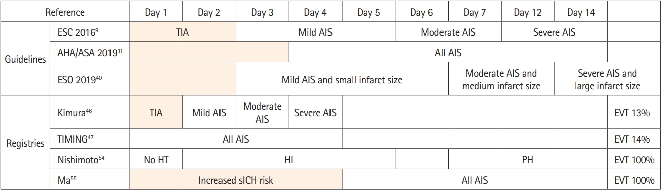
The 2016 European Society of Cardiology guidelines for the management of AF introduced the “1-3-6-12” rule for AC initiation post-AIS: 1 day after transient ischemic attack (TIA), 3 days after mild AIS (NIHSS score <8 points), 6 days after moderate AIS (NIHSS score: 8-15 points), and 12 days after severe stroke (NIHSS score >15 points) after exclusion of ICH on imaging [9]. Additional clinical and imaging factors were listed to hasten or postpone initiation of treatment, but no specific guidance was provided in case of HT or PH detection. Despite its simplicity, a drawback of a purely clinical algorithm is that it does not account for the variation in brain infarct volumes in patients with the same NIHSS score, that possibly influences HT risk. The 2016 European Stroke Organisation (ESO)-Karolinska Stroke Update underlined the importance of infarct size and proposed the “4-7-14” day rule: 4 days after mild AIS and small infarct size (lesion ≤1.5 cm), 7 days after moderate stroke and medium infarct size, and 14 days after severe AIS and large infarct size [10]. The same recommendation was included as expert opinion statement in the ESO recommendations for secondary prevention of stroke and other thromboembolic events in patients with stroke or TIA and AF, suggesting antiplatelet therapy in the first 48 hours after AF-associated stroke [24]. The American Heart Association and American Stroke Association’s guidelines are quite broad and suggest initiating AC between 4-14 days after AIS, even in the context of HT after individual assessment of risks and benefits, considering antiplatelet therapy until AC initiation [11].

Treatment individualization and risk stratification

In the absence of randomized evidence, experts suggest delaying AC in patients with severe symptomatic HT, such as PH-2 [25]. Other experts prioritize imaging findings when facing this therapeutic conundrum. They suggest initiating AC within 2 days for infarct sizes <1.5 cm in diameter, 4-5 days for sizes 1.6-3 cm at, at day 7 for infarcts 3 cm or more, 7-10 days in HT HI-1 and HI-2 and for PH-2, subarachnoid or subdural hemorrhage postponing AC for a minimum of 2-4 weeks [26]. The ALESSA score is based on patient age, lesion size, and severe atrial enlargement on transthoracic echocardiogram; higher scores are associated with high embolic risk and patients should receive earlier AC [27]. Concomitant stroke mechanisms such as cancer-related hypercoagulability, small vessel disease, and large artery atherosclerosis predicted stroke recurrence in the IAC study [28]. Therefore, it should be emphasized that an effective secondary prevention strategy post-AIS in AF patients may extend beyond AC, especially for those already on AC [29].

Safety of earlier anticoagulation in patients at high risk of recurrent embolism

An analysis of the Early Recurrence and Major Bleeding in Patients With Acute Ischemic Stroke and Atrial Fibrillation Treated With Non-Vitamin K Oral Anticoagulants (RAF-NOACs) study revealed that 5% of the patients who had initiated NOACs within 24-48 hours from index AIS developed HT, suggesting that the two days after AIS may be unsuited for AC initiation [17]. In the same analysis, the delay of 12 days to initiate AC in patients with HT as compared to those without, was not associated with a significant increase in the rate of recurrent ischemic stroke. Combined data from 2 prospective, multicenter, Japanese registries on AF patients with AIS/TIA (Stroke Acute Management with Urgent Risk-factor Assessment and Improvement [SAMURAI]-NVAF and Recurrent Embolism Lessened by rivaroxaban, an Anti-Xa agent, of Early Dosing for acute ischemic stroke and transient ischemic attack with atrial fibrillation [RELAXED]) were used to examine whether early NOAC initiation according to stroke severity was safe and effective [30]. Indeed, NOAC initiation within 1 day after TIA, within 2 days after AIS with NIHSS <8, within 3 days after AIS with NIHSS 8-15, and within 4 days after AIS with NIHSS >15 (1-2-3-4-day rule) was associated with better efficacy and similar safety compared with later initiation. Timing of Oral Anticoagulant Therapy in Acute Ischemic Stroke With Atrial Fibrillation (TIMING) is a recently published prospective, registry-based, multicenter, open-label, noninferiority, randomized-controlled trial (RCT) performed in Swedish stroke units [31]. Early NOAC initiation (≤4 days) was noninferior to late (5-10 days), as no AIS patient experienced sICH and rates of recurrent AIS and death were numerically lower in the early initiation group.

Anticoagulation after other Cardioembolic causes of stroke

AF due to moderate/severe mitral stenosis caused by rheumatic disease is considered valvular and NOACs are not formally indicated for stroke prophylaxis [32-34]. Mechanical heart valves continuously activate the coagulation cascade, leading to very high local concentrations of thrombin, meaning that very high doses of NOACs may be necessary to achieve anti-thrombin activity comparable to VKA [35-37]. As a result, VKAs are the mainstay for secondary prevention of cardioembolic stroke in patients with mechanical heart valves with an international normalized ratio (INR) goal of 3 for most stroke patients since they are considered at high risk for thromboembolism [38]. In the largest analysis of patients with mechanical heart valves presenting with acute parenchymal ICH, early re-initiation of VKA was associated with increased rates of hemorrhagic complications until 13 days after initial ICH and with respect to safety should not be routinely restarted before 14 days; the earliest day of initiation resulted day 6 reserved only for patients at high thromboembolic risk [39]. These timepoints may be seen as the maximum possible delay to initiate AC, since even AIS patients presenting with PH are believed to have a lower risk of hematoma expansion than primary ICH patients.
VKAs may also be the only AC indicated in patients with aortic or mitral bioprosthetic valves who experienced ischemic stroke under antiplatelet therapy [38]. Non-valvular AF complicated with left atrial thrombus may be seen in AIS patients; the role of NOACs is not well-established [40]. Similarly, cardioembolic stroke from left ventricular thrombus in patients with history of anterior myocardial infarction and reduced ejection fraction might necessitate VKAs for 3 months whereas the efficacy of NOACs is uncertain [40-42].

Non-cardioembolic AC indications post-stroke

Screening in patients with acute hemiplegic stroke has shown an incidence of deep vein thrombosis (DVT) of 50% within 2 weeks in the absence of heparin prophylaxis; most affect the paretic lower limb, are asymptomatic and are located below the knee [43]. A recent study showed that 5% of AIS patients treated with EVT may develop DVT and 2% may develop pulmonary embolism (PE), especially in patients with metastatic cancer [44]. The mainstay of DVT/PE treatment is AC. Compared with low-molecular weight heparin (LMWH) bridging with VKA, NOACs have been shown to be noninferior for recurrent venous thromboembolism and are associated with a lower risk of major bleeding, including intracranial bleeding [45].
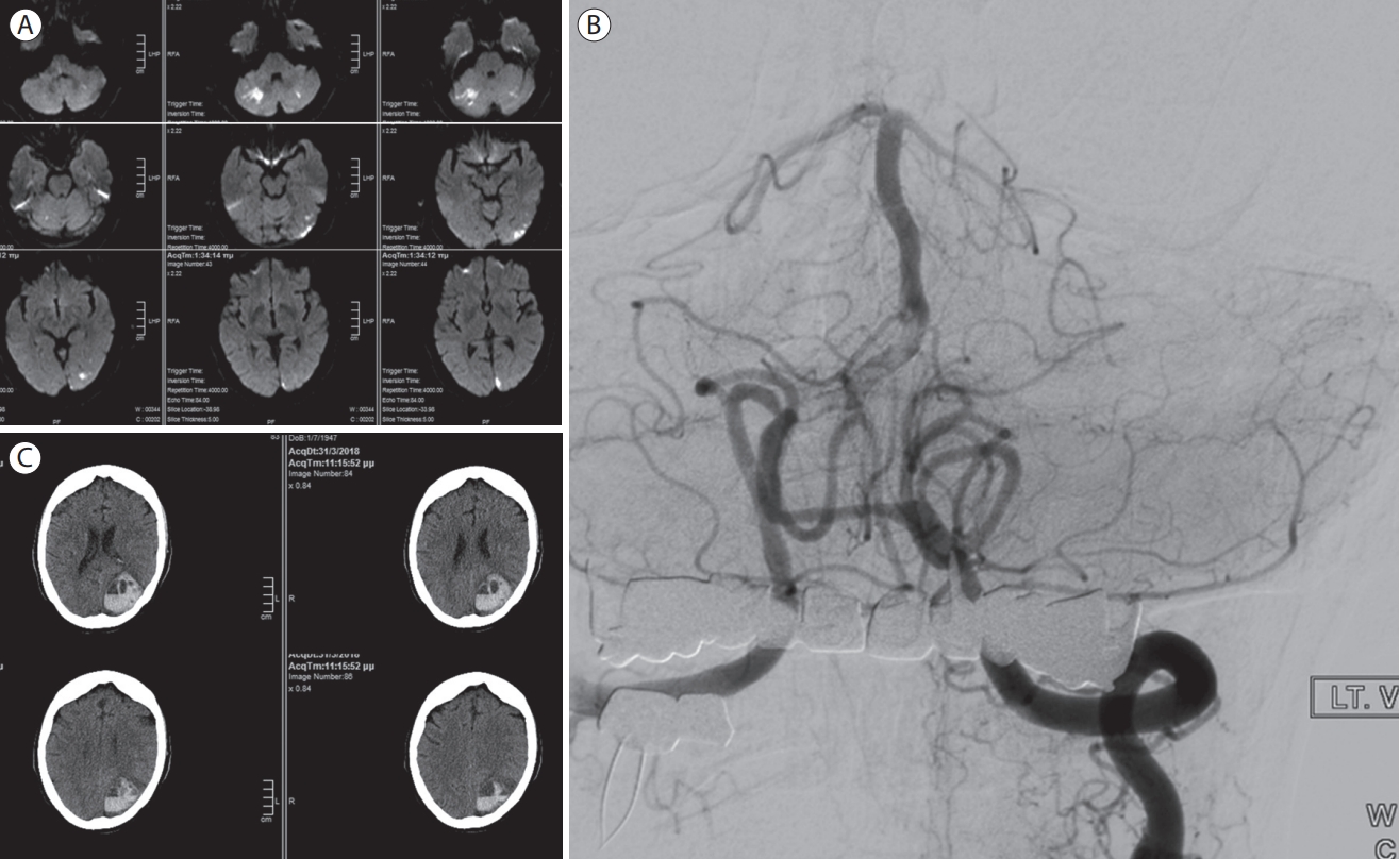
Intraluminal thrombus is detected in 1.6%-3.2% of AIS patients on computed tomography angiography, mainly due to large artery atherosclerotic disease (82%), but also caused by dissection, hypercoagulability, or cardioembolism [46]. Due to a perceived high risk of stroke recurrence, the most common antithrombotic regimen is heparin (unfractionated heparin [UFH] or LMWH) at a therapeutic dose with or without aspirin [47,48]. Figure 1 shows an illustrative case of late sICH in an AIS patient with intraluminal thrombus treated with AC following the detection of paroxysmal AF.

Recent developments

The first randomized trial on timing of NOAC anticoagulation was a study that excluded severe strokes or infarcts with ECASS grade I or II HT (NCT03433235). Patients were randomized to either a standard dose of edoxaban on day 3 for mild stroke and on day 6 for moderate stroke or half-dose of edoxaban from symptom onset until day 3 (for mild stroke) or day 6 (for moderate stroke), and a standard dose of edoxaban thereafter. This was a small trial that failed to detect any difference in clinical outcomes or late HT; however, asymptomatic ischemic lesion on MRI were numerically higher in the early initiation group, a finding that the authors attributed to the low dose of edoxaban used [49]. Early Versus Late Initiation of Direct Oral Anticoagulants in Post-ischaemic Stroke Patients With Atrial fibrillatioN (ELAN, NCT03148457) was recently published [50]. ELAN is unique in distinguishing different AIS subgroups depending on infarct size [10]. NOAC initiation within 2 days from mild or moderate infarct and within 1 week for major infarct carried the same risk for sICH (0.2%) compared with later start. There was no significant difference in recurrent stroke or systemic embolism risk, despite lower numerically rates in the early start group. These results are highly suggestive of the safety of early NOAC initiation post-stroke and are hypothesis-generating for future research regarding recurrent embolism risk reduction.

Ongoing RCTs

Three ongoing RCTs will further explore the optimal timing for AC resumption after AIS: Optimal timing of anticoagulation after acute ischemic stroke with atrial fibrillation (OPTIMAS, EudraCT, 2018003859-38) [51], Optimal Delay Time to Initiate Anticoagulation after Ischemic Stroke in Atrial Fibrillation (START, NCT03021928) [52], and Lixiana Acute Stroke Evaluation Registry (LASER, NCT03494530) [53]. Only START will include patients with HT per investigator’s judgment; LASER will exclude any PH and OPTIMAS will exclude PH-2 patients. Patients receiving systemic or endovascular reperfusion therapies will not be excluded from all three RCTs. Consequently, information to guide AC initiation in AIS patients receiving EVT that is complicated with PH is expected to remain limited in the near future.

Anticoagulation with factor XI inhibitors

Factor XI (FXI) inhibitors (asundexian and milvexian) represent the next potential generation of AC [54]. In the Program of Anticoagulation via Inhibition of FXIa by the Oral Compound BAY 2433334-NonCardioembolic Stroke (PACIFIC-STROKE) study, there was no increase in the risk of HT or ICH after early intervention and inclusion of mild and moderate stroke (NIHSS <16) and in post-intravenous thrombolysis (IVT) and/or EVT participants [55]. MRI data after study intervention showed similar incidence of HT between milvexian and placebo. Similar results were obtained in the Antithrombotic treatment with factor XIa inhibitor Milvexian to Optimize Management of Acute Thromboembolic events for Secondary Stroke Prevention (AXIOMATIC-SSP) trial [56]. Asundexian was undergoing two phase 3 RCTs (A Study to Learn How Well the Study Treatment Asundexian Works and How Safe it is Compared to Apixaban to Prevent Stroke or Systemic Embolism in People With Irregular and Often Rapid Heartbeat [Atrial Fibrillation], and at Risk for Stroke [OCEANIC-AF], NCT05643573; A multicenter, randomized, placebo-controlled, double-blind, parallel group and event driven phase 3 study of the oral FXIa inhibitor asundexian [BAY 2433334] at a dose of 50 mg od for the secondary prevention of ischemic stroke in adult patients with an acute non-cardioembolic ischemic stroke [OCEANIC-Stroke]; NCT05686070). OCEANIC-AF was stopped early due to inferior efficacy of asundexian versus apixaban [57]. Another multicenter, randomized, placebo-controlled, double-blind, parallel group and event driven phase 3 study of the oral FXIa inhibitor milvexian (BMS-986177) at a dose of 25 mg bid for the secondary prevention of ischemic stroke is conducted in adult patients with an acute non-cardioembolic ischemic stroke (LIBREXIA-Stroke; NCT05702034).

Anticoagulation before EVT

Pretreatment with therapeutic AC is a contraindication for IVT but not for EVT. Major EVT registries have shown that pretreatment with NOACs is not associated with sICH, but data are conflicting regarding VKAs. In the Bernese-European Registry for Ischemic Stroke Patients Treated Outside Current Guidelines With Neurothrombectomy Devices Using the Solitaire FR With the Intention for Thrombectomy (BEYOND-SWIFT), VKAs were associated with an increased risk of sICH and mortality, while the risk was lower with NOACs [58]. In the Spanish NORDICTUS registry, VKAs, but not NOACs, were an independent predictor of sICH [59]. In contrast, the German Stroke Registry-Endovascular Treatment (GSR-ET) failed to detect an association with AC pretreatment and early HT [60]. In conclusion, pretreatment with NOACs is consistently found safe in observational EVT studies, but more data may be needed for VKAs, since it remains possible that there exists an INR threshold beyond which hemorrhagic risk is increased. Nevertheless, current international recommendations do not advocate against EVT in large vessel occlusion patients pretreated with VKAs [11,61].

Periprocedural anticoagulation

Data from the Multicenter Randomized Clinical Trial of Endovascular Treatment of Acute Ischemic Stroke (MR CLEAN) Registry suggested that the use of IV UFH is safe during EVT [62]. In a systematic review, heparin use during EVT increased the odds of good functional outcome but carried an increased risk for sICH [63], a finding that was similar with the results of a systematic review of RCTs comparing aspirin to AC in AIS of cardioembolic origin [64]. The Multicenter Randomized Clinical Trial of Endovascular Treatment for Acute Ischemic Stroke in the Netherlands (MR CLEANMED) was an RCT that compared the combination of moderate (5,000 IU bolus followed by 1,250 IU/h for 6 h) or low-dose (5,000 IU bolus followed by 500 IU/h for 6 h) UFH with aspirin to aspirin monotherapy [65]. A significant shift towards worse functional outcomes was documented in the interim analysis for moderate-dose UFH versus no UFH; a nonsignificant shift towards the same direction has also been attested for low-dose UFH. sICH occurred more often in patients who received UFH (adjusted odds ratio 1.98; 95% confidence interval: 1.14-3.46) and the RCT was discontinued due to safety concerns. Certain major limitations of this RCT need to be acknowledged. IVT pretreatment was not a contraindication for AC, all patients received aspirin and many cases of sICH occurred in the subgroup of patients that had concurrent acute cervical carotid artery revascularization.

Bridging AC therapy

A post hoc analysis of the Early Recurrence and Cerebral Bleeding in Patients With Acute Ischemic Stroke and Atrial Fibrillation (RAF) and RAF-NOACs studies examined the efficacy and safety of bridging AC therapy. Bridging therapy was defined as full-dose LMWH started before or concurrently with VKAs, until INR reaches therapeutic range, or full-dose LMWH before initiating NOAC treatment. Bridging therapy more than doubled the odds of either ischemic or hemorrhagic outcomes and this increase was statistically significant [66]. These findings were independently reproduced in the IAC observational study that was conducted in the United States [20]. Other RCTs also confirm that bridging therapy in AF patients without AIS is either deleterious or non-beneficial during the perioperative period [67,68].

Is there any remaining indication for heparin during or after EVT?

Periprocedural single dose UFH is still being used during EVT, especially in the context of extra- or intracranial atherosclerotic disease where stenting may be performed during the emergent procedure [69,70]. UFH was used in more than a third of patients in the Basilar Artery Occlusion Endovascular Intervention versus Standard Medical Treatment (BEST) trial and its use could have been related to the increased risk of sICH associated with EVT compared to the medical treatment group (Figure 2) [71]. Intravenous glycoprotein IIb/IIIa inhibitors may be effective in preventing intraprocedural re-occlusion even if low doses may be needed to ensure safety, and clinical practice suggests that emergent use of UFH may not be necessary for stent patency but data on direct comparison with heparin are lacking [72].
A recent observational study compared ultra-early AC, defined as the initiation of UFH or LMWH <24 hours, to early AC defined as the initiation <3 days post-EVT in patients with AF. The control group was anticoagulated with VKAs, NOACs, or LMWH on or after day 4 [73]. Patients received one of two heparin dosing schemes, both without aspirin: a low-dose (generally referred to as prophylactic dose) and intermediate (initial UFH dose of 500 IU/h or maximum dose over 500 IU/h, or LMWH dose of 4,000 or 4,250 IU two times per day). Patients receiving heparin showed significantly better functional outcomes in 90 days without increase of sICH in both ultra-early and early initiation groups compared to the control group, especially in the context of ultra-early initiation which was also associated with a significant decrease in the risk of asymptomatic ICH. Whether this approach of post-procedural use of intermediate-dose UFH/LMWH is safe and effective remains to be seen in future studies.

Anticoagulation initiation after EVT

EVT does not appear to increase the risk of sICH in the Highly Effective Reperfusion evaluated in Multiple Endovascular Stroke Trials (HERMES) meta-analysis of RCTs of the anterior circulation [74]. Presumably EVT does not increase overall rates of sICH despite treatment-related hemorrhagic complications, since reducing infarct size reduces the risk of spontaneous (non-EVT-related) HT. However, in the late-time window MR CLEAN LATE trial, the risk of sICH was significantly higher in the EVT group (adjusted odds ratio 4.59) [75]. Numerically higher rates of sICH and any ICH were also noted in the ANGEL-ASPECT and Rescue Japan LIMIT randomized EVT trials of large ischemic core [76,77]. Posterior circulation EVT appears to increase sICH risk. A meta-analysis of RCTs including the recently published positive trials of basilar artery occlusion EVT showed that EVT was associated with better outcome and lower risk of death at 3 months, but a significantly higher risk of sICH was documented [78]. A similar increase in sICH has been described in isolated posterior cerebral artery (PCA) EVT although an increase in sICH was not confirmed in an isolated PCA occlusion meta-analysis comparing patients treated with EVT versus medical management [79,80]. Early HT renders early NOAC initiation problematic in patients with cardioembolic stroke.
There is scarce data on AC initiation in patients with early HT post-EVT. A recent retrospective Japanese study examined 111 patients that received EVT for anterior circulation AIS. NOACs were started at a median of 1 day in the no HT group, 3 days in the HI group (interquartile range [IQR] 2-5 days), and 7 days in the PH group (5 patients with PH-1 and 6 patients with PH-2; IQR 7-10 days) [81]. There were only 2 cases of sICH in the no HT group and 2 cases of new asymptomatic ICH in AIS patients with PH-1 occurring before initiation of NOACs; no cases of new sICH (late HT) following NOAC initiation in both HI and PH groups were documented. The study is limited by its retrospective character and its small size, with only 16 patients with HI and 13 patients with PH, but provides evidence that the risk of late HT under NOACs is low.
A prospective Chinese study suggests that very early AC initiation might be harmful. One hundred twenty-five patients requiring AC initiated treatment <5 days after EVT, 66 patients after 5-14 days and 43 patients 15 days or later after index event. sICH occurred only in the early initiation group and the difference was statistically significant, as was the increase in any ICH in the same group [82]. Moreover, EVT to AC time was an independent risk factor of sICH, ICH, and systemic hemorrhage after initiating AC. EVT for internal carotid artery occlusion was also associated with increased ICH risk after AC initiation. Embolic recurrences were similar in all treatment groups. This study provides no data on treatment strategy for patients with HT post-EVT but raises a red flag for initiating AC within the first 4 days post-EVT, especially in the context of internal carotid artery recanalization that could be related to greater infarct volume or periprocedural use of antithrombotics. A synthesis of available data with relevant expert-opinion based recommendations is provided in Figure 3 and Table 1 and a simplified illustration of the proposed treatment algorithm is available in Figure 4.

Future research

Most of the research studies that evaluate the incidence and prognostics factors of HT occurring during the first days following cardioembolic stroke with or without acute reperfusion therapies, focus on early (24-48 first hours) HT. Much less is known for late HT that is also discussed in the current narrative review. In a study on bridging AC after cardioembolic stroke, HT has been reported to occur in a bimodal distribution—an early benign and a late symptomatic PH-2—suggesting a different pathophysiology of late HT [83]. Persistent disruption of the BBB appears within 24 hours after infarction and may last for several weeks [84], but early BBB disruption may not predict delayed HT [85]. Imaging data confirms BBB derangement weeks after stroke [86]. Despite much research in developing imaging biomarkers for predicting early HT [87] and late HT [88,89], we have no reliable data on the prevalence of late intracranial bleeding (in the subacute phase of stroke) and whether AC therapy increases its risk. An elegant study using transcranial sonography to detect HT and Doppler to detect delayed recanalization indicated that time of HT development is associated with time of recanalization in patients with large vessel occlusion [90]. Moreover, HT does not always occur immediately after recanalization, but at a median delay of 40 hours. To our knowledge, this interesting observation has not been replicated. It is possible that if very late reperfusion occurs, it may lead to late HT [91]. Angiogenesis post-ischemic stroke may promote recovery but can also predispose to HT if it is premature, due to enhanced vascular permeability of newly formed, immature vessels [92].

Conclusions

Accumulating data provides valuable information on the benefits and risks of early AC after AIS. The increasing familiarity of clinicians with NOACs, which present a more favorable safety profile compared to full-dose heparin or VKAs in the subacute phase of AIS, is reflected on recently published observational studies or small phase 2 RCTs that report shorter delays to AC initiation. Given the increasing availability of EVT, more is known on AC initiation in these patients, even in the context of early HT post-recanalization. Late HT in the days and weeks following stroke remains a devastating complication for which we still do not have any reliable clinical or radiological prediction tools. Thus, it remains challenging to individualize the timepoint of AC initiation in cardioembolic stroke patients that would be associated with the lowest risk of stroke recurrence and intracranial bleeding. Among the different causes of cardioembolic stroke, non-valvular AF has been most extensively studied. We suggest that AC could be started safely in these patients 5-14 days post-stroke, depending on the risk of recurrent embolism, predicted by the ALESSA score, and the existence of early PH. More research is required to understand and predict late HT, which—in contrast to early HT—has not received attention in recent years.

Notes

Funding statement
None
Conflicts of interest
Apostolos Safouris has received a stipendium from the Hellenic Neurological Society for training in Interventional Neurology that is unrelated to the manuscript.
Author contribution
Conceptualization: AS, GT (Georgios Tsivgoulis). Study design: AS, GT (Georgios Tsivgoulis). Writing—original draft: AS, KP, LP, PO, GM, OK, GT (Georgios Tsivgoulis). Writing—review & editing: AT, TK, SS, SN, AS, TNN, SY, SW, SS, GT (Guillaume Turc). Approval of final manuscript: all authors.

Figure 1.
A 70-year-old woman, with a history of arterial hypertension and diabetes mellitus, presented with intermittent vertigo and diplopia for 48 hours. The National Institutes of Health Stroke Scale (NIHSS) on admission was 4 and magnetic resonance imaging revealed multiple acute ischemic lesions on diffusion- weighted imaging (A). Digital subtraction angiography revealed a sub-occlusive intraluminal thrombus of the right vertebral artery in the V4 segment (B). The left posterior cerebral artery was occluded in the P1 segment but slow flow to the P2 segment was noted through a small posterior communicating artery. Thrombectomy was undertaken but was unsuccessful to recanalize the right vertebral artery. The cause of intraluminal thrombus was believed to be an underlying atherosclerotic plaque, since the patient was in sinus rhythm. The patient received clopidogrel and therapeutic dose of low-molecular weight heparin (LMWH) and was discharged 4 days later with NIHSS score of 1. She presented 2 weeks after discharge with right hemianopia and aphasia (NIHSS-score: 10 points). Paroxysmal atrial fibrillation was detected on electrocardiogram. Repeat brain computed tomography revealed an extensive lobar intracranial hemorrhage of the left occipital lobe (C). LMWH and clopidogrel were halted and the patient had a neurosurgical evacuation of the hematoma with good, but partial, recovery (NIHSS score of 6 points). She was treated with apixaban 5 mg bid 12 days after surgical evacuation.
jos-2023-01578f1.jpg
Figure 2.
A 63-year-old man with atrial fibrillation presented with acute left hemianopia and gait disturbance. Emergent magnetic resonance imaging revealed an acute ischemic lesion in the distribution of the right posterior cerebral artery (PCA) (A). Digital subtraction angiography revealed a complete basilar artery (BA) occlusion (B) that was recanalized with mechanical thrombectomy (C). Distal right PCA was left occluded given the extensive established infarction of the right occipital lobe (C). The patient was loaded with aspirin (500 mg) and clopidogrel (600 mg) and received a bolus of 5,000 units of unfractionated heparin and residual BA stenosis was treated with 2 intracranial stents (D). Computed tomography (CT) post-treatment showed extravasated contrast within the right occipital lobe infarction (E). The patient had abrupt neurological deterioration 20 hours after recanalization. Emergent CT revealed an extensive parenchymal hematoma type 2 originating from the right occipital lobe (F). The patient deceased hours later.
jos-2023-01578f2.jpg
Figure 3.
Day rules proposed for initiating anticoagulation after cardioembolic stroke/TIA and registries including patients post-EVT. Days refer to days after index event as the minimal delay for each stroke category to initiate anticoagulation. Mild AIS: NIHSS <8; Moderate AIS: NIHSS 8-15; Severe AIS: NIHSS >15; Small infarct size: Ischemic lesion ≤1.5 cm; Medium infarct size: NIHSS <8 and lesion in a cortical superficial branch of middle cerebral artery (MCA), or involving the MCA deep branch, or in internal border zone territories, or in a cortical superficial branch of posterior cerebral artery (PCA), or lesion involving the PCA branch or lesion in a cortical superficial branch of anterior cerebral artery (ACA); large infarct size: anterior circulation: lesion involving complete territory of MCA, PCA, or ACA or involving 2 cortical superficial branches of MCA or involving a cortical superficial branch of MCA associated to the MCA deep branch, or involving more than 1 artery territory (eg, MCA associated to ACA territories); posterior circulation: lesion involving brain stem or cerebellum >1.5 cm. TIA, transient ischemic attack; EVT, endovascular treatment; ESC, European Society of Cardiology; AIS, acute ischemic stroke; AHA/ASA, American Heart Association and American Stroke Association; ESO, European Stroke Organisation; HT, hemorrhagic transformation; HI, hemorrhagic infarction; PH, parenchymal hemorrhage; NIHSS, National Institutes of Health Stroke Scale.
jos-2023-01578f3.jpg
Figure 4.
Synthesis of available data for initiating anticoagulation (AC) after endovascular treatment (EVT). Starting non-vitamin K antagonist oral anticoagulants (NOACs) without bridging is possible at day 2 but may be safer at day 5, even in the context of hemorrhagic infarction. Delaying initiation at 7-14 days for patients with parenchymal hemorrhage may be safe. Aspirin should be given until NOAC initiation. ALESSA scores 3-4 should prompt earlier AC initiation.
jos-2023-01578f4.jpg
Table 1.
Individualized treatment strategy for initiating anticoagulation in acute ischemic stroke patients with atrial fibrillation receiving EVT
Study Treatment strategy
van der Steen et al. [65] Avoid unfractionated heparin during EVT
Altavilla et al. [66] Do not bridge oral anticoagulation with full dose heparin
Palaiodimou et al. [22] Prefer NOACs
Ma et al. [82] Starting oral anticoagulation >4 days from symptom onset may reduce intracranial hemorrhage risk
Paciaroni et al. [27] Consider earlier initiation of oral anticoagulation in patients with ALESSA* scores 3-4
Nishimoto et al. [81] - Starting oral anticoagulation ≤14 days from symptom onset may reduce risk of recurrent ischemic stroke
- Hemorrhagic infarction: initiation <7 days from symptom onset appears safe (earliest time point of initiation: 5 days)
- Parenchymal hemorrhage: initiation ≤14 days from symptom onset appears safe (earliest time point of initiation: 7 days)
EVT, endovascular treatment; NOACs, non-vitamin K antagonist oral anticoagulants.
* ALESSA score (0-4 points): 1 point for patient age 70-79, 2 points for those 80 or older, 1 point for ischemic lesion on neuroimaging with diameter >1.5 cm, and 1 point for severe atrial enlargement on transthoracic echocardiogram.

References

1. Aguilar MI, Hart R, Pearce LA. Oral anticoagulants versus antiplatelet therapy for preventing stroke in patients with non-valvular atrial fibrillation and no history of stroke or transient ischemic attacks. Cochrane Database Syst Rev 2007;3:CD006186.
crossref
2. Liu C, Shi F, Chen Z, Yan S, Ding X, Lou M. Severe blood-brain barrier disruption in cardioembolic stroke. Front Neurol 2018;9:55.
crossref pmid pmc
3. Kobeissi H, Ghozy S, Seymour T, Gupta R, Bilgin C, Kadirvel R, et al. Outcomes of patients with atrial fibrillation following thrombectomy for stroke: a systematic review and meta-analysis. JAMA Netw Open 2023;6:e2249993.
crossref pmid pmc
4. Berge E, Abdelnoor M, Nakstad PH, Sandset PM. Low molecular-weight heparin versus aspirin in patients with acute ischaemic stroke and atrial fibrillation: a double-blind randomised study. Lancet 2000;355:1205-1210.
crossref pmid
5. Saxena R, Lewis S, Berge E, Sandercock PA, Koudstaal PJ. Risk of early death and recurrent stroke and effect of heparin in 3169 patients with acute ischemic stroke and atrial fibrillation in the international stroke trial. Stroke 2001;32:2333-2337.
crossref pmid
6. Fang MC, Go AS, Chang Y, Borowsky LH, Pomernacki NK, Udaltsova N, et al. Thirty-day mortality after ischemic stroke and intracranial hemorrhage in patients with atrial fibrillation on and off anticoagulants. Stroke 2012;43:1795-1799.
crossref pmid pmc
7. Paciaroni M, Agnelli G, Corea F, Ageno W, Alberti A, Lanari A, et al. Early hemorrhagic transformation of brain infarction: rate, predictive factors, and influence on clinical outcome: results of a prospective multicenter study. Stroke 2008;39:2249-2256.
crossref pmid
8. van der Steen W, van der Ende NA, van Kranendonk KR, Chalos V, Brouwer J, van Oostenbrugge RJ, et al. Timing of symptomatic intracranial hemorrhage after endovascular stroke treatment. Eur Stroke J 2022;7:393-401.
crossref pmid pmc pdf
9. Kirchhof P, Benussi S, Kotecha D, Ahlsson A, Atar D, Casadei B, et al. 2016 ESC guidelines for the management of atrial fibrillation developed in collaboration with EACTS. Eur J Cardiothorac Surg 2016;50:e1-e88.
pmid
10. Ahmed N, Steiner T, Caso V, Wahlgren N; ESO-KSU Session Participants. Recommendations from the ESO-Karolinska Stroke Update Conference, Stockholm 13-15 November 2016. Eur Stroke J 2017;2:95-102.
crossref pmid pmc pdf
11. Powers WJ, Rabinstein AA, Ackerson T, Adeoye OM, Bambakidis NC, Becker K, et al. Guidelines for the early management of patients with acute ischemic stroke: 2019 update to the 2018 guidelines for the early management of acute ischemic stroke: a guideline for healthcare professionals from the American Heart Association/American Stroke Association. Stroke 2019;50:e344-e418.
pmid
12. Nogueira RG, Gupta R, Jovin TG, Levy EI, Liebeskind DS, Zaidat OO, et al. Predictors and clinical relevance of hemorrhagic transformation after endovascular therapy for anterior circulation large vessel occlusion strokes: a multicenter retrospective analysis of 1122 patients. J Neurointerv Surg 2015;7:16-21.
crossref pmid pmc
13. Hacke W, Kaste M, Fieschi C, von Kummer R, Davalos A, Meier D, et al. Randomised double-blind placebo-controlled trial of thrombolytic therapy with intravenous alteplase in acute ischaemic stroke (ECASS II). Lancet 1998;352:1245-1251.
crossref pmid
14. Hacke W, Kaste M, Bluhmki E, Brozman M, Dávalos A, Guidetti D, et al. Thrombolysis with alteplase 3 to 4.5 hours after acute ischemic stroke. N Engl J Med 2008;359:1317-1329.
crossref pmid
15. Wahlgren N, Ahmed N, Dávalos A, Ford GA, Grond M, Hacke W, et al. Thrombolysis with alteplase for acute ischaemic stroke in the Safe Implementation of Thrombolysis in Stroke-Monitoring Study (SITS-MOST): an observational study. Lancet 2007;369:275-282.
crossref pmid
16. von Kummer R, Broderick JP, Campbell BC, Demchuk A, Goyal M, Hill MD, et al. The Heidelberg bleeding classification: classification of bleeding events after ischemic stroke and reperfusion therapy. Stroke 2015;46:2981-2986.
crossref pmid
17. Paciaroni M, Bandini F, Agnelli G, Tsivgoulis G, Yaghi S, Furie KL, et al. Hemorrhagic transformation in patients with acute ischemic stroke and atrial fibrillation: time to initiation of oral anticoagulant therapy and outcomes. J Am Heart Assoc 2018;7:e010133.
pmid pmc
18. Tsivgoulis G, Katsanos AH, Seiffge DJ, Paciaroni M, Wilson D, Koga M, et al. Fatal intracranial haemorrhage occurring after oral anticoagulant treatment initiation for secondary stroke prevention in patients with atrial fibrillation. Eur J Neurol 2020;27:1612-1617.
crossref pmid pdf
19. Seiffge DJ, Paciaroni M, Wilson D, Koga M, Macha K, Cappellari M, et al. Direct oral anticoagulants versus vitamin K antagonists after recent ischemic stroke in patients with atrial fibrillation. Ann Neurol 2019;85:823-834.
pmid pmc
20. Yaghi S, Mistry E, Liberman AL, Giles J, Asad SD, Liu A, et al. Anticoagulation type and early recurrence in cardioembolic stroke: the IAC study. Stroke 2020;51:2724-2732.
crossref pmid pmc
21. Hong KS, Kwon SU, Lee SH, Lee JS, Kim YJ, Song TJ, et al. Rivaroxaban vs warfarin sodium in the ultra-early period after atrial fibrillation-related mild ischemic stroke: a randomized clinical trial. JAMA Neurol 2017;74:1206-1215.
crossref pmid pmc
22. Palaiodimou L, Stefanou MI, Katsanos AH, Paciaroni M, Sacco S, De Marchis GM, et al. Early anticoagulation in patients with acute ischemic stroke due to atrial fibrillation: a systematic review and meta-analysis. J Clin Med 2022;11:4981.
crossref pmid pmc
23. Polymeris AA, Zietz A, Schaub F, Meya L, Traenka C, Thilemann S, et al. Once versus twice daily direct oral anticoagulants in patients with recent stroke and atrial fibrillation. Eur Stroke J 2022;7:221-229.
crossref pmid pmc pdf
24. Klijn CJ, Paciaroni M, Berge E, Korompoki E, Kõrv J, Lal A, et al. Antithrombotic treatment for secondary prevention of stroke and other thromboembolic events in patients with stroke or transient ischemic attack and non-valvular atrial fibrillation: a European Stroke Organisation guideline. Eur Stroke J 2019;4:198-223.
crossref pmid pmc pdf
25. Seiffge DJ, Werring DJ, Paciaroni M, Dawson J, Warach S, Milling TJ, et al. Timing of anticoagulation after recent ischaemic stroke in patients with atrial fibrillation. Lancet Neurol 2019;18:117-126.
crossref pmid
26. Malhotra K, Khunger M, Liebeskind DS. Non-vitamin K oral anticoagulants in stroke patients: practical issues. J Stroke 2017;19:104-106.
crossref pmid pmc pdf
27. Paciaroni M, Agnelli G, Caso V, Tsivgoulis G, Furie KL, Tadi P, et al. Prediction of early recurrent thromboembolic event and major bleeding in patients with acute stroke and atrial fibrillation by a risk stratification schema: the ALESSA score study. Stroke 2017;48:726-732.
pmid
28. Yaghi S, Henninger N, Giles JA, Leon Guerrero C, Mistry E, Liberman AL, et al. Ischaemic stroke on anticoagulation therapy and early recurrence in acute cardioembolic stroke: the IAC study. J Neurol Neurosurg Psychiatry 2021;92:1062-1067.
crossref pmid pmc
29. Yaghi S. Diagnosis and management of cardioembolic stroke. Continuum (Minneap Minn) 2023;29:462-485.
crossref pmid
30. Kimura S, Toyoda K, Yoshimura S, Minematsu K, Yasaka M, Paciaroni M, et al. Practical “1-2-3-4-day” rule for starting direct oral anticoagulants after ischemic stroke with atrial fibrillation: combined hospital-based cohort study. Stroke 2022;53:1540-1549.
pmid pmc
31. Oldgren J, Åsberg S, Hijazi Z, Wester P, Bertilsson M, Norrving B; National TIMING Collaborators. Early versus delayed non-vitamin K antagonist oral anticoagulant therapy after acute ischemic stroke in atrial fibrillation (TIMING): a registry-based randomized controlled noninferiority study. Circulation 2022;146:1056-1066.
crossref pmid pmc
32. Steffel J, Collins R, Antz M, Cornu P, Desteghe L, Haeusler KG, et al. 2021 European Heart Rhythm Association practical guide on the use of non-vitamin K antagonist oral anticoagulants in patients with atrial fibrillation. Europace 2021;23:1612-1676.
pmid
33. Connolly SJ, Karthikeyan G, Ntsekhe M, Haileamlak A, El Sayed A, El Ghamrawy A, et al. Rivaroxaban in rheumatic heart disease-associated atrial fibrillation. N Engl J Med 2022;387:978-988.
pmid
34. Eikelboom JW, Connolly SJ, Brueckmann M, Granger CB, Kappetein AP, Mack MJ, et al. Dabigatran versus warfarin in patients with mechanical heart valves. N Engl J Med 2013;369:1206-1214.
crossref pmid
35. Fanaroff AC, Vora AN, Lopes RD. Non-vitamin K antagonist oral anticoagulants in patients with valvular heart disease. Eur Heart J Suppl 2022;24(Suppl A):A19-A31.
crossref pmid pmc pdf
36. Jawitz OK, Wang TY, Lopes RD, Chavez A, Boyer B, Kim H, et al. Rationale and design of PROACT Xa: a randomized, multicenter, open-label, clinical trial to evaluate the efficacy and safety of apixaban versus warfarin in patients with a mechanical On-X aortic heart valve. Am Heart J 2020;227:91-99.
crossref pmid pmc
37. Neale T. Another DOAC fails in the setting of mechanical heart valves [Internet]. New York: TCTMD; 2022 September 28 [accessed May 6, 2023]. Available from: https://www.tctmd.com/news/another-doac-fails-setting-mechanical-heart-valves.

38. Otto CM, Nishimura RA, Bonow RO, Carabello BA, Erwin JP 3rd, Gentile F, et al. 2020 ACC/AHA guideline for the management of patients with valvular heart disease: a report of the American College of Cardiology/American Heart Association Joint Committee on clinical practice guidelines. Circulation 2021;143:e72-e227.
pmid
39. Kuramatsu JB, Sembill JA, Gerner ST, Sprügel MI, Hagen M, Roeder SS, et al. Management of therapeutic anticoagulation in patients with intracerebral haemorrhage and mechanical heart valves. Eur Heart J 2018;39:1709-1723.
pmid pmc
40. Kleindorfer DO, Towfighi A, Chaturvedi S, Cockroft KM, Gutierrez J, Lombardi-Hill D, et al. 2021 guideline for the prevention of stroke in patients with stroke and transient ischemic attack: a guideline from the American Heart Association/American Stroke Association. Stroke 2021;52:e364-e467.
pmid
41. Robinson AA, Trankle CR, Eubanks G, Schumann C, Thompson P, Wallace RL, et al. Off-label use of direct oral anticoagulants compared with warfarin for left ventricular thrombi. JAMA Cardiol 2020;5:685-692.
crossref pmid pmc
42. Huang L, Tan Y, Pan Y. Systematic review of efficacy of direct oral anticoagulants and vitamin K antagonists in left ventricular thrombus. ESC Heart Fail 2022;9:3519-3532.
crossref pmid pmc pdf
43. Kelly J, Rudd A, Lewis R, Hunt BJ. Venous thromboembolism after acute stroke. Stroke 2001;32:262-267.
crossref pmid
44. Aboul-Nour H, Maraey A, Jumah A, Khalil M, Elzanaty AM, Elsharnoby H, et al. Mechanical thrombectomy for acute ischemic stroke in metastatic cancer patients: a nationwide cross-sectional analysis. J Stroke 2023;25:119-125.
crossref pmid pmc pdf
45. Gómez-Outes A, Lecumberri R, Suárez-Gea ML, Terleira-Fernández AI, Monreal M, Vargas-Castrillón E. Case fatality rates of recurrent thromboembolism and bleeding in patients receiving direct oral anticoagulants for the initial and extended treatment of venous thromboembolism: a systematic review. J Cardiovasc Pharmacol Ther 2015;20:490-500.
crossref pmid pdf
46. Psychogios K, Kargiotis O, Safouris A, Repousis P, Tsivgoulis G. Floating carotid thrombus in a patient with essential thrombocytemia due to calreticulin mutation. Stroke 2023;54:e207-e208.
crossref pmid
47. Singh RJ, Chakraborty D, Dey S, Ganesh A, Al Sultan AS, Eesa M, et al. Intraluminal thrombi in the cervico-cephalic arteries. Stroke 2019;50:357-364.
crossref pmid
48. Das S, Goldstein ED, de Havenon A, Abbasi M, Nguyen TN, Aguiar de Sousa D, et al. Composition, treatment, and outcomes by radiologically defined thrombus characteristics in acute ischemic stroke. Stroke 2023;54:1685-1694.
crossref pmid
49. Kim JS, Lee KB, Chang DI, Cha JK, Lee JS. Early administration of edoxaban after acute ischemic stroke in patients with non-valvular atrial fibrillation: a pilot randomized trial. J Stroke 2023;25:311-314.
crossref pmid pmc pdf
50. Fischer U, Koga M, Strbian D, Branca M, Abend S, Trelle S, et al. Early versus later anticoagulation for stroke with atrial fibrillation. N Engl J Med 2023;388:2411-2421.
pmid
51. Best JG, Arram L, Ahmed N, Balogun M, Bennett K, Bordea E, et al. Optimal timing of anticoagulation after acute ischemic stroke with atrial fibrillation (OPTIMAS): protocol for a randomized controlled trial. Int J Stroke 2022;17:583-589.
crossref pmid pdf
52. King BT, Lawrence PD, Milling TJ, Warach SJ. Optimal delay time to initiate anticoagulation after ischemic stroke in atrial fibrillation (START): methodology of a pragmatic, response-adaptive, prospective randomized clinical trial. Int J Stroke 2019;14:977-982.
crossref pmid pmc pdf
53. Alrohimi A, Jickling G, Jeerakathil T, Shuaib A, Khan K, Kate M, et al. Protocol for LASER: a randomized evaluation and an associated registry of early anticoagulation with edoxaban after ischemic stroke in patients with atrial fibrillation. Front Neurol 2021;12:645822.
crossref pmid pmc
54. Georgi B, Mielke J, Chaffin M, Khera AV, Gelis L, Mundl H, et al. Leveraging human genetics to estimate clinical risk reductions achievable by inhibiting factor XI. Stroke 2019;50:3004-3012.
crossref pmid pmc
55. Shoamanesh A, Mundl H, Smith EE, Masjuan J, Milanov I, Hirano T, et al. Factor XIa inhibition with asundexian after acute non-cardioembolic ischaemic stroke (PACIFIC-stroke): an international, randomised, double-blind, placebo-controlled, phase 2b trial. Lancet 2022;400:997-1007.
crossref pmid
56. Sharma M, Molina CA, Toyoda K, Bereczki D, Bangdiwala SI, Kasner SE, et al. Safety and efficacy of factor XIa inhibition with milvexian for secondary stroke prevention (AXIOMATIC-SSP): a phase 2, international, randomised, double-blind, placebo-controlled, dose-finding trial. Lancet Neurol 2024;23:46-59.
crossref pmid
57. Bayer. OCEANIC-AF study stopped early due to lack of efficacy [Internet]. Leverkusen: Bayer; 2023 November 19 [accessed November 29, 2023]. Available from: https://www.bayer.com/media/en-us/oceanic-af-study-stopped-early-due-to-lack-of-efficacy/.

58. Meinel TR, Kniepert JU, Seiffge DJ, Gralla J, Jung S, Auer E, et al. Endovascular stroke treatment and risk of intracranial hemorrhage in anticoagulated patients. Stroke 2020;51:892-898.
crossref pmid
59. Ramos-Araque ME, Chavarría-Miranda A, Gómez-Vicente B, López-Cancio Martínez E, Castañón Apilánez M, Castellanos M, et al. Oral anticoagulation and risk of symptomatic hemorrhagic transformation in stroke patients treated with mechanical thrombectomy: data from the nordictus registry. Front Neurol 2020;11:594251.
crossref pmid pmc
60. Küpper C, Feil K, Wollenweber FA, Tiedt S, Herzberg M, Dorn F, et al. Endovascular stroke treatment in orally anticoagulated patients: an analysis from the German stroke registry-endovascular treatment. J Neurol 2021;268:1762-1769.
crossref pmid pmc pdf
61. Turc G, Bhogal P, Fischer U, Khatri P, Lobotesis K, Mazighi M, et al. European Stroke Organisation (ESO) - European Society for Minimally Invasive Neurological Therapy (ESMINT) guidelines on mechanical thrombectomy in acute ischaemic strokeendorsed by Stroke Alliance for Europe (SAFE). Eur Stroke J 2019;4:6-12.
crossref pmid pmc pdf
62. van de Graaf RA, Chalos V, van Es ACGM, Emmer BJ, Lycklama À Nijeholt GJ, van der Worp HB, et al. Periprocedural intravenous heparin during endovascular treatment for ischemic stroke: results from the MR CLEAN registry. Stroke 2019;50:2147-2155.
pmid pmc
63. van de Graaf RA, Chalos V, Del Zoppo GJ, van der Lugt A, Dippel DWJ, Roozenbeek B. Periprocedural antithrombotic treatment during acute mechanical thrombectomy for ischemic stroke: a systematic review. Front Neurol 2018;9:238.
pmid pmc
64. Paciaroni M, Agnelli G, Micheli S, Caso V. Efficacy and safety of anticoagulant treatment in acute cardioembolic stroke: a meta-analysis of randomized controlled trials. Stroke 2007;38:423-430.
crossref pmid
65. van der Steen W, van de Graaf RA, Chalos V, Lingsma HF, van Doormaal PJ, Coutinho JM, et al. Safety and efficacy of aspirin, unfractionated heparin, both, or neither during endovascular stroke treatment (MR CLEAN-MED): an open-label, multicentre, randomised controlled trial. Lancet 2022;399:1059-1069.
crossref pmid
66. Altavilla R, Caso V, Bandini F, Agnelli G, Tsivgoulis G, Yaghi S, et al. Anticoagulation after stroke in patients with atrial fibrillation. Stroke 2019;50:2093-2100.
pmid
67. Douketis JD, Spyropoulos AC, Kaatz S, Becker RC, Caprini JA, Dunn AS, et al. Perioperative bridging anticoagulation in patients with atrial fibrillation. N Engl J Med 2015;373:823-833.
crossref pmid pmc
68. Kovacs MJ, Wells PS, Anderson DR, Lazo-Langner A, Kearon C, Bates SM, et al. Postoperative low molecular weight heparin bridging treatment for patients at high risk of arterial thromboembolism (PERIOP2): double blind randomised controlled trial. BMJ 2021;373:n1205.
crossref pmid pmc
69. Yang M, Huo X, Gao F, Wang A, Ma N, Liebeskind DS, et al. Safety and efficacy of heparinization during mechanical thrombectomy in acute ischemic stroke. Front Neurol 2019;10:299.
crossref pmid pmc
70. Lee JS, Lee SJ, Hong JM, Alverne FJAM, Lima FO, Nogueira RG. Endovascular treatment of large vessel occlusion strokes due to intracranial atherosclerotic disease. J Stroke 2022;24:3-20.
crossref pmid pmc pdf
71. Liu X, Dai Q, Ye R, Zi W, Liu Y, Wang H, et al. Endovascular treatment versus standard medical treatment for vertebrobasilar artery occlusion (BEST): an open-label, randomised controlled trial. Lancet Neurol 2020;19:115-122.
pmid
72. Zhu X, Cao G. Safety of glycoprotein IIb-IIIa inhibitors used in stroke-related treatment: a systematic review and meta-analysis. Clin Appl Thromb Hemost 2020;26:1076029620942594.
crossref pmid pmc pdf
73. Xu Y, Liu C, Li W, Nie X, Huang S, Li X, et al. Efficacy and safety of early anticoagulation after endovascular treatment in patients with atrial fibrillation. Stroke Vasc Neurol 2023;8:405-412.
crossref pmid pmc
74. Goyal M, Menon BK, van Zwam WH, Dippel DW, Mitchell PJ, Demchuk AM, et al. Endovascular thrombectomy after large-vessel ischaemic stroke: a meta-analysis of individual patient data from five randomised trials. Lancet 2016;387:1723-1731.
crossref pmid
75. Olthuis SGH, Pirson FAV, Pinckaers FME, Hinsenveld WH, Nieboer D, Ceulemans A, et al. Endovascular treatment versus no endovascular treatment after 6-24 h in patients with ischaemic stroke and collateral flow on CT angiography (MR CLEAN-LATE) in the Netherlands: a multicentre, open-label, blinded-endpoint, randomised, controlled, phase 3 trial. Lancet 2023;401:1371-1380.
pmid
76. Huo X, Ma G, Tong X, Zhang X, Pan Y, Nguyen TN, et al. Trial of endovascular therapy for acute ischemic stroke with large infarct. N Engl J Med 2023;388:1272-1283.
pmid
77. Yoshimura S, Sakai N, Yamagami H, Uchida K, Beppu M, Toyoda K, et al. Endovascular therapy for acute stroke with a large ischemic region. N Engl J Med 2022;386:1303-1313.
crossref pmid
78. Palaiodimou L, Eleftheriou A, Katsanos AH, Safouris A, Magoufis G, Spiliopoulos S, et al. Endovascular treatment for acute basilar artery occlusion: a fragility index meta-analysis. J Clin Med 2023;12:2617.
crossref pmid pmc
79. Sabben C, Charbonneau F, Delvoye F, Strambo D, Heldner MR, Ong E, et al. Endovascular therapy or medical management alone for isolated posterior cerebral artery occlusion: a multicenter study. Stroke 2023;54:928-937.
pmid
80. Berberich A, Finitsis S, Strambo D, Michel P, Herweh C, Meyer L, et al. Endovascular therapy versus no endovascular therapy in patients receiving best medical management for acute isolated occlusion of the posterior cerebral artery: a systematic review and meta-analysis. Eur J Neurol 2022;29:2664-2673.
pmid
81. Nishimoto T, Oka F, Okazaki K, Ishihara H. Timing of direct oral anticoagulants for hemorrhagic transformation after endovascular treatment in acute ischemic stroke. J Stroke Cerebrovasc Dis 2022;31:106507.
crossref pmid
82. Ma H, Che R, Zhang Q, Yu W, Wu L, Zhao W, et al. The optimum anticoagulation time after endovascular thrombectomy for atrial fibrillation-related large vessel occlusion stroke: a real-world study. J Neurol 2023;270:2084-2095.
crossref pmid pmc pdf
83. Hallevi H, Albright KC, Martin-Schild S, Barreto AD, Savitz SI, Escobar MA, et al. Anticoagulation after cardioembolic stroke: to bridge or not to bridge? Arch Neurol 2008;65:1169-1173.
crossref pmid pmc
84. Strbian D, Durukan A, Pitkonen M, Marinkovic I, Tatlisumak E, Pedrono E, et al. The blood-brain barrier is continuously open for several weeks following transient focal cerebral ischemia. Neuroscience 2008;153:175-181.
crossref pmid
85. Jickling GC, Liu D, Stamova B, Ander BP, Zhan X, Lu A, et al. Hemorrhagic transformation after ischemic stroke in animals and humans. J Cereb Blood Flow Metab 2014;34:185-199.
crossref pmid pmc pdf
86. Liu HS, Chung HW, Chou MC, Liou M, Wang CY, Kao HW, et al. Effects of microvascular permeability changes on contrast-enhanced T1 and pharmacokinetic MR imagings after ischemia. Stroke 2013;44:1872-1877.
crossref pmid
87. Elsaid N, Mustafa W, Saied A. Radiological predictors of hemorrhagic transformation after acute ischemic stroke: an evidence-based analysis. Neuroradiol J 2020;33:118-133.
crossref pmid pmc pdf
88. Wilson D, Ambler G, Shakeshaft C, Brown MM, Charidimou A, Al-Shahi Salman R, et al. Cerebral microbleeds and intracranial haemorrhage risk in patients anticoagulated for atrial fibrillation after acute ischaemic stroke or transient ischaemic attack (CROMIS-2): a multicentre observational cohort study. Lancet Neurol 2018;17:539-547.
pmid pmc
89. Martí-Fàbregas J, Medrano-Martorell S, Merino E, Prats-Sánchez L, Marín R, Delgado-Mederos R, et al. MRI predicts intracranial hemorrhage in patients who receive long-term oral anticoagulation. Neurology 2019;92:e2432-e2443.
pmid pmc
90. Seidel G, Cangür H, Albers T, Burgemeister A, Meyer-Wiethe K. Sonographic evaluation of hemorrhagic transformation and arterial recanalization in acute hemispheric ischemic stroke. Stroke 2009;40:119-123.
crossref pmid
91. Bernardo-Castro S, Sousa JA, Brás A, Cecília C, Rodrigues B, Almendra L, et al. Pathophysiology of blood-brain barrier permeability throughout the different stages of ischemic stroke and its implication on hemorrhagic transformation and recovery. Front Neurol 2020;11:594672.
crossref pmid pmc
92. Rust R. Insights into the dual role of angiogenesis following stroke. J Cereb Blood Flow Metab 2020;40:1167-1171.
crossref pmid pmc pdf
TOOLS
Share :
Facebook Twitter Linked In Google+
Graph View
  • 0 Crossref
  •   Scopus
  • 4,127 View
  • 204 Download


ABOUT JoS
AUTHOR INFORMATION
ARTICLE CATEGORY

Browse all articles >

BROWSE ARTICLES
Editorial Office
Department of Neurology, Asan Medical Center,Ulsan University College of Medicine
88, Olympic-ro 43-gil, Songpa-gu, Seoul 05505, Korea
Submission, status and progress, etc ⟫ E-mail: editor@j-stroke.org
Website and system ⟫ E-mail: journal@m2community.co.kr
Publishing company ⟫ E-mail: ka72sus@smileml.com
Developed in M2PI
Copyright © 2024 by Korean Stroke Society.
Close layer
prev next